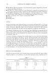
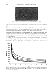
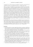
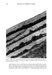
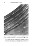
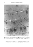
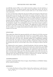
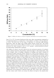
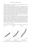
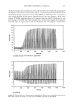
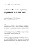
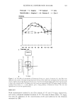
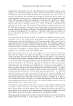
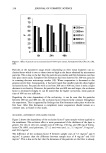
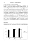
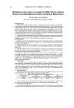
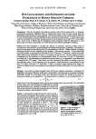
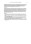
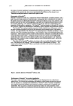
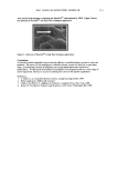
268 JOURNAL OF COSMETIC SCIENCE .. Figure 2. Cross section of normal hair pretreated with uranyl acetate/lead citrate. The stains have pen- etrated the fiber, giving contrast to the endocuticle (•--), cell membrane complex (•n) and structures in the cortex. Scale bar = 0.5 pm. nitrate staining. In the cuticle the a-layer and exocuticle showed intense staining. Sparse staining was seen in the endocuticle. None of the component structures of the cmc were easily observed. Localized deposits of silver were seen within the cell membrane complex of the cuticle (Figures 4, 5). The high-sulphur proteins of the

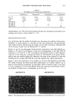
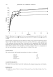
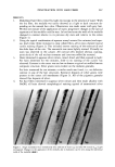
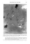
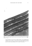
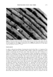
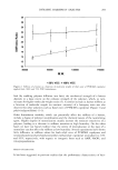
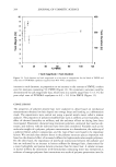
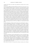
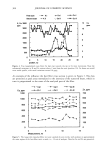
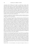
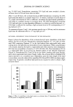
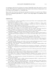
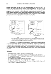
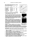
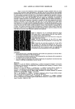
PENETRATION INTO HAIR FIBER 269 Figure 3. Pretreatment with aqueous silver nitrate, after which the hair was rinsed, dried, and exposed to light. Note the extensive silver deposits in the cortical membranes (•,•). There are pigment granules ((in) but no apparent contrast in the macrofibrils. Scale bar = 0.3 lain. 5(i). cortex were intensely stained, revealing a typical microfibril/matrix ultrastructure. Localized deposits of silver were seen within the cell membrane complex of the cortex and within some inter-macrofibrillar material. For hair immersed in sodium chloride (two minutes), dried, followed by immer-
Purchased for the exclusive use of nofirst nolast (unknown)
From: SCC Media Library & Resource Center (library.scconline.org)