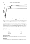
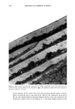
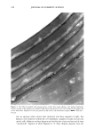
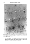
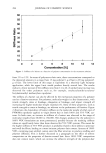
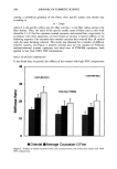
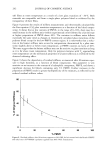
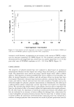
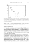
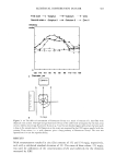
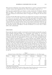
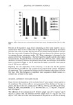
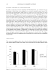
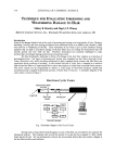
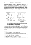
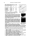
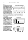
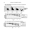
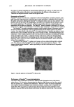
ELEMENTAL DISTRIBUTION IN HAIR 309 forensic examinations. In the present study a variation of the concentrations for elements in all hairs was recorded. To study the reproducibility of our method we scanned along a hair with high K content (Figure 4). As can be seen, the two scans correspond well for K and Ca variations, while as for Cu with its concentration around 10 mg/g, the reproducibility is somewhat lower. Apart from variations in time in the stability of the instrument, positioning of the sample, etc., another possible but very minor source of error in this experiment could be variations in hair length due to changes in air hu- midity. The difference in length between a dry and a wet fiber is not much more than 1%. A strong rise in the concentration of K occurs some 3-4 mm from the root. This corresponds approximately to where the fiber has lost the outer root sheath, i.e., is exposed to the environment and thus effectively is brought above the skin surface. It is then exposed to the excretion of the skin glands and to the environment, and hence all data from measurements in that part of the hair are likely to suffer from contamination effects. Another aspect of reproducibility is the observation that the standard deviation for zinc in the present study is much lower than in the preceeding PIXE study based on the same material (9). Earlier studies indicate that zinc has a relatively even distribution over the cross section, while the sulfur concentration varies. This can partly explain the discrepancy in measured sulfur values between the studies, as the data in the PIXE-based study primarily arise from the hair surface. The sulfur content of the intact exocuticle has been estimated to be about 11% (25). Selecting the empirical median value from each group of hairs (each person) and exclu- sively including these data in the database make it more likely that occasional errors have been excluded. One type of problem that might be solved by this means is the alteration of analytical data by particles occurring on the hair surface. In the hair groups, i.e., the three or six hairs from each person, there is, however, a tendency of co-variation. Thus, if a particular fiber was shown to be rich in a certain element, other hairs of the same sample group were also high in this element, as can be seen when comparing mean and median data (Tables I and II). This correlation has not been studied in detail. Monovalent ions appear to be relatively loosely bound to the hair fiber material. As was mentioned above, the K concentration can be expected to vary a lot between different sites on a fiber (Figure 4). The monovalent ions K and CI are highly water-soluble and are easily washed out when cleansing the hair in distilled or preferably in deionized water. When surfactants are used, they should be of the non-ionic type for obvious reasons. To a certain extent, this is also true for Ca, but Ca is sometimes present in tap water in considerable amounts and can thus be absorbed also. CONCLUSIONS The present investigation introduces a new energy-dispersive X-ray technique for el- emental analysis of biological materials, here represented by hair fibers. The method proves to have distinct advantages over particle probes in that it causes no loss of material during the analyses, with the doses used. This results in data corresponding to "live concentrations" at a level of sensitivity that matches that of PIXE (proton-induced X-ray emission) analysis. The particle probes produce characteristic secondary X-ray emission when electrons or protons are retarded or stopped by elements in the specimen.

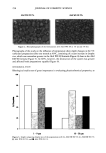
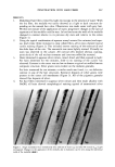
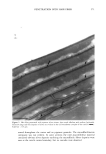
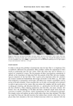
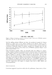
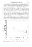
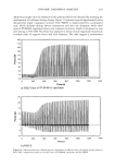
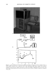
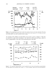
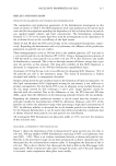
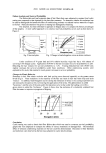
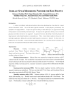
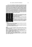
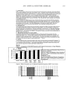
310 JOURNAL OF COSMETIC SCIENCE A secondary effect of this is that the specimen suffers mass loss due to heating in the volume analyzed, which is not the case in X-ray fluorescence analysis. Furthermore, reexamination of the same section (volume) of the specimen is possible with our XRF instrumentation. The reproducibility is highly satisfactory, taking into account the precision with which the actual sampled volume can be reestablished and the biological variation of elemental concentrations as a function of position in the sample. X-ray fluorescence spectrometry is a sensitive bulk method. However, this property is less pronounced for low X-ray energies (light elements), due to attenuation of the emitted low-energy photons in the bulk material. The effect is clearly demonstrated in the experiments using hair fiber rotation (Figure 3). The variation in hair fiber diameter is less pronounced for the heavy metals such as Zn than for the light elements such as S and K. The elemental concentrations recorded in this study are in reasonable agreement with those obtained earlier (9), taking into account that the previous PIXE data were not corrected for variations in the cross sections. These data will provide a basis for future analysis of pathological hair fibers and for forensic analysis of hair fibers retrieved in crime investigations. ACKNOWLEDGMENTS We are indebted to the National Laboratory of Forensic Science of Sweden for making resources available for this study, and to the Edvard Welander and Finsen Foundations, the research funds of the Karolinska Institutet, for financial support. REFERENCES (1) J. Andrasko and B. Stocklassa, Shampoo residue profiles in human hair, J. Forensic Sci., 35, 596-579 (1990). (2) A.J.J. Bos, On the incorporation of trace elements into human hair measured with micro-PIXE, Nuclear Instr, Meth. Phys. Res, B3, 654-659 (1984). (3) O. Braun-Falco and E. Christoffers "Hair Root Pattern in Male Pattern Alopecia," in Biopatho/ogy of Pattern Alopecia, A. Baccaredda-Boy, G Moretti, and JR Frey, Eds. (Karger, Basel and New York, 1968), pp. 141-145. (4) W. R. Collie, T. J. Goka, C. M. Moore, and R. R. Howe, "Hair in Menke's Disease: A Comprehensive Review," in Hair, Trace Elements and Human Illness, Brown and Crounse, Eds. (Praeger Scientific, New York, 1980), pp. 197-209. (5) D.J. Eatough, J.J. Christensen, R. M. Izatt, and C. Hartley, "Levels of Selected Trace Elements in Human Hair," in The First Human Hair Symposium, A. C. Brown, Ed. (Medcom Press, New York, 1974), pp. 377-387. (6) P. Engstr/Sm, Development of Capillary Optics for X-ray Focusing, Thesis, Chalmers Institute of Technol- ogy, Gothenburg, Sweden, 1991. (7) P. Engstr/Sm, S. Larsson, A. Rindby, and B. Stocklassa, A 200 rnm X-ray microbeam spectrometer, Nucl. Instr. Meth. Phys. Res., B36, 222-226 (1989). (8) B. Forslind, "The Growing Ariagert Hair," in Hair and Hair Diseases, C. E. Orfanos and R. Happie, Eds. (Springer Verlag, Berlin, New York, London, Paris, Tokyo, 1990), pp. 73-97. (9) B. Forslind, H. K. Li, K. G. Malmqvist, and D. Wiegleb, Elemental content of ariagert hairs in a normal Caucasian population studies with proton induced X-ray emission (PIXE), Scanning Electron Microsc., I, 237-241 (1986). (10) B. Forslind, K. Wiren, and K. G. Malmqvist, Assessment of qualitative and quantitative data from
Purchased for the exclusive use of nofirst nolast (unknown)
From: SCC Media Library & Resource Center (library.scconline.org)