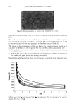
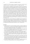
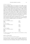
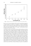
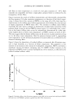
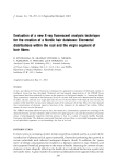
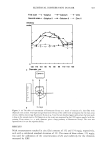
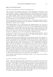
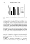
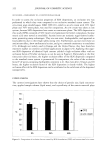
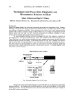
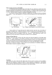
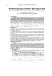
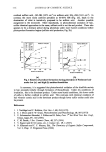
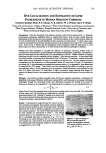
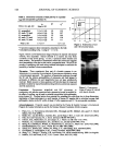
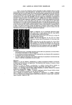
ELEMENTAL DISTRIBUTION IN HAIR 299 amounts of hair, making them practically useless for analysis of virgin parts of hair. In forensic applications and studies of certain medical disorders, such amounts are not available and single-fiber analysis becomes necessary. This type of analysis has until now only been possible with particle probes and synchrotron light (2,9,10,18,22-24), which allows the analysis of very small volumes, making it possible to reanalyze the same part of the specimen. We will demonstrate that the X-ray fluorescence (XRF) technique presented here has the advantage of being highly sensitive at trace-element levels of detection and that, in addition, it is a non-destructive technique. In the present study we have investigated triplets of anagen (actively growing) hair fibers collected from the temporal area of 63 normal Caucasians, using XRF analysis in a new energy-dispersive analysis system. It was not possible to obtain a blood sample simul- taneously with the collection of hair fibers. MATERIALS AND METHODS THE INSTRUMENT ITRAX •, is a new commercially available X-ray spectrometer used for the analyses (Figure 1). The ITRAX system is useful for bulk and trace element analysis in small samples, using X-Ray fluorescence (XRF) analysis. The detection limits depend on the photon energy emitted from the excited sample by a particular element and reach down to single tag/g for some elements in hair (Figure 2). The principles of the instrument have been previously described (6,7,15,16,21). Briefly, the X-ray source, which can be op- erated at a maximum of 60kV/50mA, is an X-ray tube with a molybdenum (Mo) anode. The X-rays are transmitted to the sample through a conical glass capillary that has diameters of 0.5 mm at the end facing the anode and 0.3 mm at the outer end, allowing for a bremsstrahlung reduction and an intensity increase by concentration of the X-rays on the specimen through subsequent multiple reflections inside the capillary. The divergency of the beam at the exit is less than 0.2 ø . The measuring area of the instrument is protected by a plastic hood, and it is operated in air. In the particular setup for hair analysis, the position of the sample can be visualized through two CCD cameras equipped with lenses focused on the object. One camera faces the sample in a direction opposite to the extension of the impinging X-ray beam the other camera operates from above. These cameras are connected to a video monitor which also allows measurement of the fiber cross section seen by the camera as well as the position of the beam seen by using a fluorescent screen placed in the beam. The fluorescent and scattered X-rays were registered using a 30-mm 2 energy-dispersive Si(Li) detector with a Link 2040 pulse processor placed at a 90 ø angle to the beam pulse processor. The detector of the specific equipment used has 146 eV resolution at 5.9 keV and is connected to a Camberra-Packard multi-channel analyzer installed in a Win- dowsT•4-based PC. Spectra were evaluated using a spectrum evaluation program (Spetest, Cox Analytical) Cox Analytical Systems AB, Teatergatan 36, 411 35 Gothenburg, Sweden.

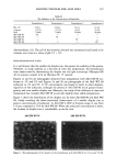
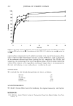
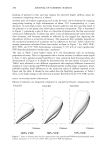
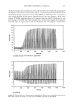
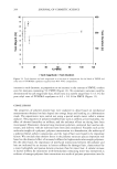
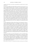
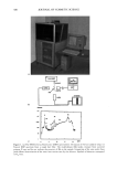
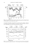
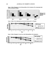
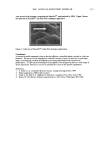
300 JOURNAL OF COSMETIC SCIENCE C ß ß Jl '•s •a keV Figure 1. (a) The ITRAX X-ray fluorescence (XRF) spectrometer. (b) Layout of the hair analysis setup. (c) Typical XRF spectrum from a single hair fiber. The molybdenum (Mo) peaks emanate from scattered primary X-rays and do not indicate the presence of Mo in the sample. Integration of the area under these peaks allows determination of the mass cross section seen by the detector. Symbols of elements correspond to K,, lines.
Purchased for the exclusive use of nofirst nolast (unknown)
From: SCC Media Library & Resource Center (library.scconline.org)