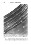
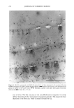
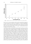
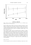
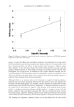
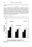
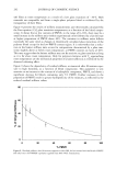
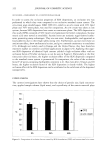
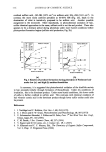
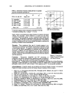
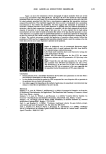
2001 ANNUAL SCIENTIFIC SEMINAR 343 contains glycerin, which absorbs more water when in contact'with liquid water and, therefore, becomes more permeable than petrolatum. Water vapor transport through different thicknesses of vernix was also investigated. The correlation between permeability coefficient and the reciprocal of thickness was consistent with Fick's first law of diffusion. Vernix water vapor permeability also showed marked temperature dependence in the physiological range from 25øC to 37 Conclusions The data are consistent with the hypothesis that vernix has a skin protective role before and after birth. The temperature dependence of the rheology shows that vernix will spread in utero but will be difficult to remove post parturn when the skin surface cools. While vemix is permeable to water and allows vapor transport it presents a hydrophobic surface that repels water. These data indicate that vernix has important differences from other commonly used skin creams. This information is critical to formulating bioengineered material films based on vernix as a prototype. Further characterization of this naturally occurring biofilm may allow insight into the mechanism(s) by which the fetus develops a functionally mature and highly aesthetic skin surface. References 1. W.L. Pickens, R.R. Warner, R.E. Boissy, and S.B. Hoath. Characterization of vernix caseosa: water content, morphology, and elemental analysis. Journal of Investigative Dermatology 115: 875-881, 2000. 2. Y. ,Sumida, M. Yakumaru, Yo Tokitsu, Y. Iwamato, T. Ikemoto, and K. Mimura. Studies on the function of vernix caseosa, the secrecy of baby's skin. Preprints of the IFSCC 20th International Conference, Cannes, France Poster 201, 1998. 3. W. Youssef, R.R. Wickett, and S.B. Hoath. Surface free energy characterization of vernix caseosa. Potential role in waterproofing the newborn infant. Skin Research and Technology 7:10-17, 2001. 4. A. Martin. Rheology. In'. Physical Pharmacy, edited by A. Martin. 453-476, 1993 5. M. Walker, T. Hulme, M. Rippon, R. Walmsley, ,S. Gunnigle, M. Lewin, and S. Winsey. In vitro model(s) for the percutaneous delivery of active tissue repair agents. Journal of Pharmaceutical Sciences 86: 1379-1384, 1997.

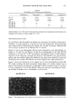
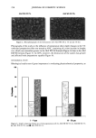
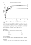
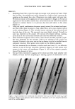
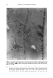
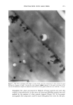
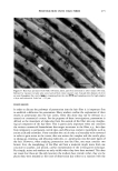
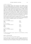
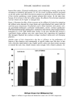
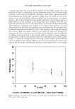
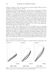
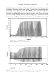
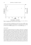
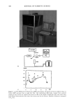
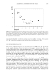
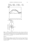
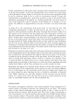
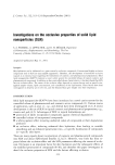
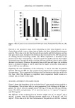
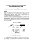
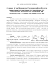
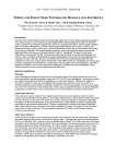
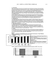
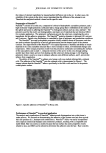
344 JOURNAL OF COSMETIC SCIENCE LIPID NANOTUBES AS SKIN PENETRATION MODULATORS Vitthal Kulkarni, Ph.D., Joretta Wong, Duncan Aust, Ph.D., James Wilmott and James Hayward, Ph.D. The Collaborative Group, Ltd., 3 Technology Drive, East Setauket, NY 11733 vitt haL kulkarni @ c ollabo. com INTRODUCTION Human skin is constantly exposed to the harsh environment including bright sun light, atmospheric pollutants and extreme wet or dry conditions that cause premature skin aging and may lead to derreal conditions. Topical supplement of skin care actives is necessary to maintain a healthy skin. To get the actives safely into skin there needs to be a delivery system that can carry the actives efficiently and be non toxic to skin. Advantages of a suitable "delivery system" include the possibility of reduced toxicity, controlled release, and deep penetration of an active into skin (Kulkami et al., 2001 a, 2001b). In cosmetic formulations it is equally important to retard or prohibit the penetration of actives such as sunscreens. Sphingolipids are an important class of lipids that have unusual thermotropic properties than the normal glycerol based phospholipids and play very important role in structure and function of cell membranes. Recent investigation has revealed that certain sphingolipids including cerebrosides and ceramides form tubular microstructures upon hydration (Kulkami et al. 1999). However, the tubular microstructures have found limited application in the delivery systems (Zarif et al. 2000a 2000b). An appropriate combination of phospholipids, sphingolipids and certain other lipids form tubular microstructures that have shown promising applications in the topical delivery systems as penetration enhancers. Our recent studies indicate that depending on the mode of application of nanotubes, they can promote or retard skin penetration significantly. Here we report our studies on lipid nanotubes and their applications in cosmetic delivery systems as controllers of skin penetration. MATERIALS AND METHODS Lipid nanotubes were formed with proprietary high-pressure high-shear technique. Retinol palmitate was incorporated into the tubes as an active ingredient. A nano-dispersion of retinol palmitate was also prepared with established procedure. A panel of five volunteers was chosen to test the products "Nanotubes retinol palmitate" and "nano-dispersion retinol palmitate" both at the same concentration (0.5%). Known amount of test product was applied to the forearm of volunteers at four different places. At desired time points after application, the applied surface area was rinsed with ethanol to recover un- penetrated active. Retinol palmitate from skin washings was quantitatively analyzed via HPLC. In a separate study we applied a solution of nanotubes for 30 min (at 37øC) to a model skin epidermis equivalent to human skin, (Epi 606 MatTek, Inc.) followed by incubation with a nano-dispersion of oil which also contained a fluorescent dye octadecyl filuorescene (ODF) for 2 hrs at 37øC. The model skin was then examined in a confocal microscope to determine the depth of penetration of the marker dye ODF. RESULTS AND DISCUSSION * '• b•r = 1 un• , , 0 • 4 6 8 "Hn'•, he Figure 1: A: Transmission electron micrograph of lipid nanotubes. B: Retinol palmitate penetrated deep into skin. Curve I represents nanotube formulation and curve 2 represents the nano-dispersion
Purchased for the exclusive use of nofirst nolast (unknown)
From: SCC Media Library & Resource Center (library.scconline.org)