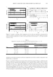
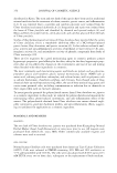
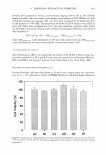
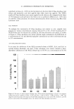
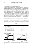
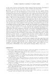
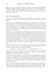
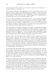
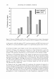
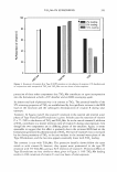
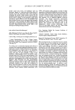
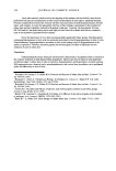
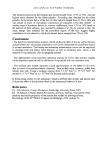
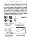
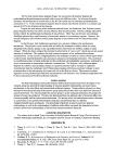
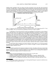
U. DA VIDIANA EXTRACTS IN COSMETICS 361 Samples were prepared at various concentrations ranging from 0.1 % to 3%. Normal human fibroblast cells were treated with samples in the absence of 10% FBS for 4 h, after which the medium was changed. The cells were then incubated for an additional 20 h in the presence of 10% FBS. Ten microliters of thawed CCK-8 solution was added to each well. Plates were incubated for 4 h at the same incubator conditions, after which the absorbance was read at 450 nm with a reference wavelength of 600 nm. Cell viability was calculated as: Cell viability (%) = (OD4SO(sample ) / OD4SO(conrrol ) ) X 100 where OD 450(sample) is the absorbance at 450 nm of the treated cells and OD 45 o(concrol) is the absorbance at 450 nm of the negative control (non-treated cells). ANTI-INFLAMMATORY EFFECTS Anti-inflammatory effects were quantified by means of the ELISA method using com mercially available kits. 11-6 and 11-8 were detected using kits from Endogen (Woburn, MA), and PGE2 was detected with kits from Assay Design Inc. (Ann Arbor, Ml). RECOVERY OF PHOTO-INDUCED DAMAGE (13,14) Human fibroblast cells were distributed to 24-well tissue culture plates at a concentra tion of 2.5 x 10⁵ cells/well in 500 µl of DMEM (Dulbecco's Modified Eagle's Medium) 120 100 80 - :.a 60 40 20 0 (a) (b) (c) (d) (e) (f) Figure 4. Cytotoxicity of U Imus davidiana root extract against human fibroblast cells: (a) control (b) 0.1 % (c) 0.5% (d) 1 % (e) 2% (f) 3%. The data are expressed as mean values (± standard deviations) of four experiments.

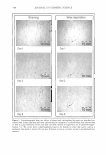
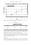
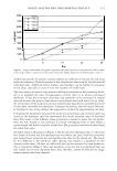
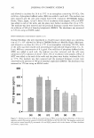
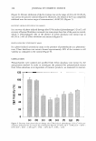
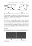
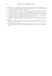
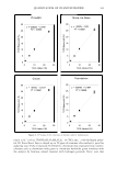
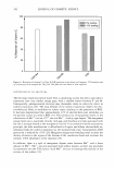
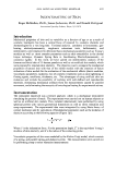
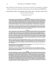
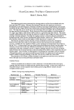
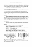
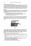
362 JOURNAL OF COSMETIC SCIENCE and allowed to incubate for 24 h at 37°C in an atmosphere containing 5% CO2• One milliliter of phosphate-buffered saline (PBS) was added to each well. The medium was then removed and the cells were treated with UV A irradiation (F30T8BLB, Sankyo Denki, Tokyo, Japan, 3 J/cm2 ). After 24 h of incubation with samples, 100 µl of MTT was added to each of the wells, and the plates were further incubated for 4 h at 3 7°C. The medium was then removed and the produced formazan crystals were dissolved by the addition of 500 µl of dimethyl sulphoxide (DMSO). The absorbance was measured at 5 70 nm using an ELISA reader. PHOTO-INDUCED CYTOTOXICITY ASSAY (13,14) Human fibroblast cells were distributed to 24-well tissue culture plates at a concentra tion of 2.5 x 10⁵ cells/well in 500 µl of DMEM (Dulbecco's Modified Eagle's Medium) and allowed to incubate for 24 h at 37°C in an atmosphere containing 5% CO 2 • Each of the wells was then treated with promethazine hydrochloride (Sigma Chem. Co., St. Louis, MO) at a concentration of 0.5 µg/ml. One milliliter of phosphate-buffered saline (PBS) was added to each well, the medium was then removed, and the cells were subjected to UV irradiation (3 J/cm2). After 24 h of incubation with samples, 100 µl of MTT was added to the each of the wells and the plates were further incubated for 4 h at 37 ° C. The medium was then removed and the produced formazan crystals were dissolved by the addition of 500 µI of dimethyl sulphoxide (DMSO). The absorbance was measured at 570 nm using an ELISA reader. 500 - 400 -- 300 Q) Q) Q) 200 L.. N UJ C) a.. 100 0 (a) (b) (c) (d) (e) (f) Figure 5. Inhibitory activity of H2O2-activated release of PEG2 in normal human fibroblast cell lines: (a) negative control (b) positive control (c) 0.01 % (d) 0.025% (e) 0.05% (f) 0.1 %. The negative control and the positive control were H2Oruntreated cells and H 2 O2-treated cells in the absence of U Imus davidiana root extract, respectively.
Purchased for the exclusive use of nofirst nolast (unknown)
From: SCC Media Library & Resource Center (library.scconline.org)