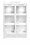
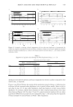
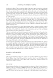
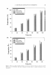
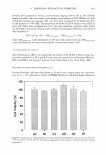
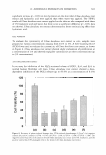
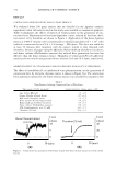
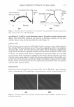
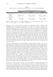
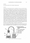
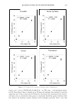
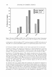
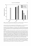
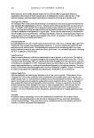
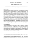
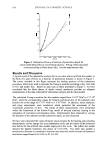
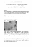
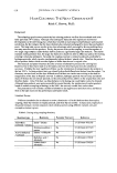
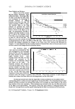
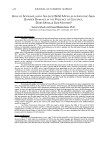
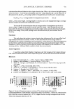
374 JOURNAL OF COSMETIC SCIENCE Table II Efficacy of Gels, Including the Horse Chestnut Extract, on Facial-Wrinkle Smoothing Wrinkle score (mean ± SE) 0 week 6 weeks 9 weeks Corner of eye Sample 0.766 ± 0.104 0.597** ± 0.082 0.613** ± 0.100 Placebo 0.895 ± 0.139 0.956 ± 0.120 0.975## ± 0.121 Lower eyelid Sample 1.20 ± 0.083 1.03** ± 0.089 0.95*** ± 0.089 Placebo 1.24 ± 0.094 1.25 ± 0.102 1.19# ± 0.089 Each value represents the mean ± SE. Significantly different from corresponding 0-week values: **p 0.01, ***p p 0.001 (sample) #p 0.05, ##p 0.001 (placebo). Therefore, active contraction forces generated by fibroblasts appear to influence the morphology and/or mechanical properties of the skin in one way or another. Few ingredients that generate cell contraction forces are known except LP A or thrombin and the like (1). Though these ingredients have been shown to regulate various biological activities in vitro, very little is known of these effects in vivo (6). Moreover, it is difficult to use those ingredients for cosmetic or external use because of their cost and/or safety. Therefore, we searched among various plant extracts for ingredients that generate cell contraction forces using a dermal equivalent model. We found that an extract of horse chestnuts (Aesculus hippocastanum) is able to generate such contraction forces in fibro blasts. We found several other active plant extracts in the same screening. No plant extract that generates cell contraction force had been reported previously therefore, these results show the first discovery of a plant extract that has the activity to generate cell contraction force. The horse chestnut extract is the most effective among the plant extracts tested, and so we examined the mechanism of force generation by this extract as a representative case. The involvement of actin polymerization is suggested by the fact that cytochalasin D (an inhibitor of actin polymerization) inhibited the force generation. In addition, the for mation of stress fibers was observed in fibroblasts treated with this extract. These results suggest that the horse chestnut extract induces contraction force by the formation of stress fibers accompanied by actin polymerization. It has been reported that force gen eration by thrombin or LP A is due to phosphorylation of the myosin light chain (7). Although further study is required, the above-mentioned results indicate that phos phorylation of the myosin light chain and the formation of stress fibers are important in the force generation of cells by horse chestnut extract. The mechanisms of action of other plant extracts were not investigated. However, it can be speculated that a similar action occurs, since similar force/time-dependent curves were observed following treatment with horse chestnut extract and other effective extracts. Fortunately, the horse chestnut extract is safe and suitable for cosmetic formulation, and therefore we evaluated the in vivo efficacy of this extract on wrinkle smoothing as a representative anti-aging effect on skin morphology. Clinical tests were carried out using simple gel formulations that included 3% of the horse chestnut extract or a placebo. The gels were applied topically to the periphery of the eye skin at least two times daily for nine weeks. The horse chestnut extract showed a significant wrinkle-smoothing efficacy

HORSE CHESTNUT EXTRACT VS SKIN AGING 375 at the corner of the eye and the lower eyelid compared with the placebo. Six weeks of treatment with the gel was sufficient to have a wrinkle-smoothing effect. Medically, it is well known that extracts of horse chestnut are active against chronic venous insufficiency and anti-inflammation activities (8,9). But these biological and physiological activities were not explained due to the contraction force generation. Here, we show the wrinkle-smoothing effect of this extract. We hypothesize that the genera tion of contraction forces by fibroblasts, followed by a firming of the dermis, causes this efficacy. The activity of the ingredients to generate cell contraction forces in the skin in vivo is not clear, but cell contraction forces play important roles not only in vitro but also in vivo. Berg et al. (10) have reported that cytochalasin D induces edema formation and the lowering of interstitial fluid pressure in the dermis. Their results suggest that dermal cells can participate in the regulation of the extracellular matrix fluid of tissues via the actin filament system, and that dynamic assembly and disassembly of actin filaments also occur in the cells of dermal tissue in vivo. In general, the elastic properties of the skin change in edema. Auriol et al. (11) have reported that immediate extensibility, Ue, which reflects the elastic properties of the skin, is decreased in lymphoedema. Moreover, there have been several studies that indicate the negative relationship between the elasticity of the skin and wrinkle formation (12,13). Taken together, it is quite likely that the disruption of the actin filament system induces skin edema followed by wrinkle formation. To the contrary, it is suggested that the extract of horse chestnut acts upon the actin filament system and generates cell contraction force, resulting in wrinkle smoothing efficacy accompanied by contraction of dermal tissue. We did not test this extract in a controlled study compared with other ingredients such as ascorbic acid (14) or alpha-lipoic acid (15), and we did not clarify the mechanism(s) of wrinkle smoothing experimentally in vivo. Although further investigation will be required, our results suggest that the horse chestnut extract, which induces the con traction force of fibroblasts in vitro! exerts potent anti-aging efficacy even in vivo. REFERENCES (1) M. S. Kolodney and R. B. Wysolmerski, Isometric contraction by fibroblasts and endothelial cells in tissue culture: A quantitative study, J. Cell Biol., 117, 73-82 (1992). (2) A. K. Harris, P. Wild, and D. Stopak, Silicone rubber substrata: A new wrinkle in the study of cell locomotion, Science, 208, 177-179 (1980). (3) N. M. Morel, P. P. Petruzzo, H.B. Hechtman, and D. Shepro, Inflammatory agonises that increase microvascular permeability in vivo stimulate cultured pulmonary microvessel endothelial cell contrac tion, Inflammation, 14, 571-583 (1990). (4) E. Bell, B. Ivarsson, and C. Merrill, Production of a tissue-like structure by contraction of collagen lattices by human fibroblasts of different proliferative potential in vitro, Proc. Natl. Acad. Sci. U.S.A., 76, 1274-1278 (1979). (5) K. Tsukahara, Y. Takema, H. Kazama, Y. Yorimoto, T. Fujimura, S., Moriwaki, T. Kitahara, M. Kawai, and G. Imokawa. A photographic scale for the assessment of human facial wrinkles,]. Cosmet. Sci., 51, 127-139 (2000). (6) L. Balazs, J. Okolicany, M. Ferrebee, B. Tolley, and G. Tigyi, Topical application of the phospholipid growth factor lysophosphatidic acid promotes wound healing in vivo, Am. J. Physiol. Regul. lntegr. Comp. Physiol., 280, R466-R472 (2001). (7) M. S. Kolodney and E. L. Elson, Correlation of myosin light chain phosphorylation with isometric contraction of fibroblasts,]. Biol. Chem., 268, 23850-23855 (1993). (8) M. H. Pittler and E. Ernst, Horse-chestnut extract for chronic venous insufficiency: A criteria-based systematic review, Arch. Dermatol., 134, 1356-1360 (1998).
Purchased for the exclusive use of nofirst nolast (unknown)
From: SCC Media Library & Resource Center (library.scconline.org)