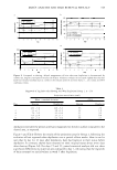
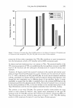
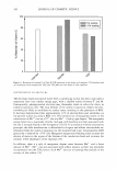
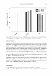
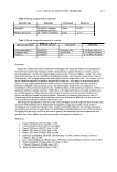
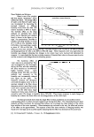
U. DA VIVIANA EXTRACTS IN COSMETICS 367 izing effect. HPLC experiments showed that the main components of the polysaccharide are rhamnose and galactose. U lmus davidiana root extract showed nearly the same mois turizing effect as hyaluronic acid. In an anti-inflammatory effect assay for inhibition of the H2Oractivated release of PGE2, IL-6, and IL-8 in normal human fibroblast cell lines, U lmus davidiana root extract showed inhibitory activity of PGE2 production in a dose-dependent manner (up to 85 .9% at a concentration of 0.1 % ). Percent inhibition of the release of IL-6 was in the range of 45.6-64.5% (H202 was used as the positive control). Moreover, the release of IL-8 was completely inhibited over the entire range of concentration (0.0025%). In a photo-induced cytotoxicity assay in the presence of promethazine as a photosensitizer, Ulmus davidiana root extract showed approximately 48% of the increase in cell viability as compared to the control. It also demonstrated good recovery from UV A-induced damage. Therefore, U lmus davidiana root extract has potential for future use as a cosmetic ingredient. ACKNOWLEDGMENT This work was supported by a grant from the Ministry of Health and Welfare: "New Material Research Center for Cosmeceuticals" (Grant Number 2005-A050432). REFERENCES (1) S. J. Lee, Korea Folk Medicine (Publishing Center of Seoul National University, Seoul, South Korea, 1996), monographs series No. 3, pp. 39-42. (2) C. D.Jun, H. 0. Pae, Y. C. Kim, S. J.Jeong,J. C. Yoo, E. J. Lee, B. M. Choi, S. W. Choe, R. K. Park, and H. T. Chung, Inhibition of nitric oxide synthesis by butanol fraction of the methanol extract of Ulmus davidiana in murine macrophages,]. Ethnopharmacol., 62, 129-135 (1998). (3) B. W. Son, J. H. Park, and 0. P. Zee, Catechin glycoside from Ulmus davidiana, Arch. Phann. Res., 12, 219-222 (1989). (4) S. H. Kim, K. T. Hwang, and J. C. Park, Isolation of flavonoids and determination of rutin from the leaves of Ulmus jJarvifolia, Kor. J. Pharmacogn., 23, 229-234 (1992). (5) Y. H. Moon and G. R. Rim, Studies on the constituents of Ulmusparvifolia, Kor.]. Pharmcogn., 26, 1-7 (1995). (6) E. B. Lee, 0. K. Kim, C. S. Jung, and K. H. Jung, The influence of methanol extract of Ulmus davidiana var. japonica cortex on gastric erosion and ulcer and paw edema in rats,]. Pharrn. Soc. Korea, 39, 671-675 (1995). (7) N. D. Hong, Y. S. Rho, N. J. Kim, and J. S. Kim, A study on efficacy of Ulmi cortex, Kor. J. Pharmacogn., 21, 217-222 (1990). (8) S. K. Cho, S. G. Lee, and C. J. Kim, Anti-inflammatory and analgesic activities of water extract of root bark of Ulmus parvifolia, Kor. J. Pharmacogn., 27, 274-281 (1996). (9) Y. M. Yang, J. W. Hyun, K. H. Lim, M. S. Sung, S.S.Kang, W. H. Paik, K. W. Bae, H. Cho, H.J. Kim, E. R. Woo, H.K. Park, and J. G. Park, Antineoplastic effect of extracts from traditional medicine plants and various plants (III), Kor. J. Pharmacogn., 27, 105-110 (1996). (10) Y. M. Kwon, J. H. Lee, and M. W. Lee, Phenolic compounds from barks of Ulmus macrocarpa and its antioxidative activities, Kor. J. Pharmacogn., 33, 404-410 (2002). (11) G. R. Leonardi, L. R. Gaspar, and P. M. B. G. Maia Campos, Application of a non-invasive method to study the moisturizing effect of formulations containing vitamins A or E or ceramide on human skin, J. Cosmet. Sci., 53, 263-268 (2002). (12) J. Levin and H. Maibach, The correlation between transepidermal water loss and percutaneous ab sorption: An overview,]. Controlled Release, 105, 291-299 (2005). (13) M. Richard, R. Lasarow, I. Rivkah, and C. G. Edward, Quantitative in vitro assessment of phototox icity by a fibroblast-neutral red assay, j. Invest. Dermatol. 98, 725-729 (1992). (14) K. B. Eberlein, A. Bindl, and B. Przybilla, Phototoxic properties of neuroleptic drugs, Dermatology, 194, 131-135 (1997).

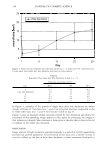
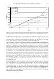
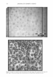
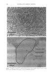
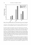
J. Cosmet. Sci., 57, 369-376 (September/October 2006) A horse chestnut extract, which induces contraction forces in fibroblasts, is a potent anti-aging ingredient TSUTOMU FUJIMURA, KAZUE TSUKAHARA, SHIGERU MORIWAKI, MITSUYUKI HOTTA, TAKASHI KITAHARA, and YOSHINORI TAKEMA, Biological Science Laboratories, Kao Corporation, 2606, Akabane, Ichikai-rnachi, Haga-gun, Tochigi, 321-3497, Japan. Accepted for publication May 4, 2006. Synopsis Contraction forces generated by non-muscle cells, such as fibroblasts, play important roles in determining cell morphology, vasoconstriction, and/or wound healing. We have searched among various plant extracts for ingredients that generate cell contraction forces using fibroblast-populated collagen gels. Using that model, we found that an extract of horse chestnuts (Aesculus hippocastanum) is able to generate such con traction forces in fibroblasts. The involvement of stress fiber formation in that response is suggested by the inhibition of such force generation by cytochalasin D and rhodamine phalloidin stain. Clinical testing of the extract was carried out using 40 healthy female volunteers. A gel formulation that included 3% of the extract was applied topically to the skin around the eye three times daily for nine weeks. The efficacy of the extract to diminish wrinkles was evaluated by visual scoring based on photo scales. After six weeks, significant decreases in the wrinkle scores at the corners of the eye or in the lower eyelid skin were observed compared with controls. After nine weeks, similar resLilts were obtained. Taken together, our results suggest that an extract of horse chestnuts can generate contraction forces in fibroblasts and is a potent anti-aging ingredient. INTRODUCTION Contraction forces generated by non-muscle cells, such as fibroblasts, play important roles in determining cell morphology, vasoconstriction, and/or wound healing (1-3). In general, non-muscle cells are thought to generate from 1/10 to 1/100 the force of smooth muscle cells, which specialize in generating such forces (1 ). Therefore, active contraction forces generated by fibroblasts appear to influence the morphology and/or mechanical properties of the skin. However, ingredients that generate cell contraction forces are hardly known except for lysophosphatidic acid (LP A), thrombin, or fetal bovine serum and the like (1,3). Address all correspondence to Tsutomu Fujimura.
Purchased for the exclusive use of nofirst nolast (unknown)
From: SCC Media Library & Resource Center (library.scconline.org)