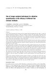
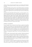
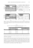
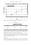
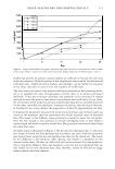
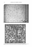
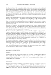
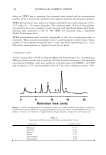
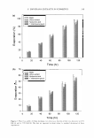
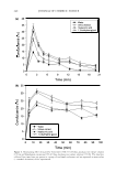
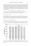
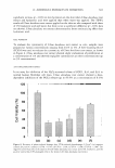
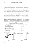
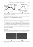
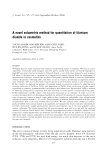
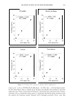
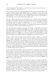
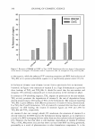
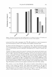
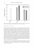
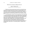
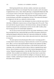
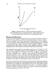
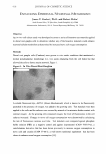
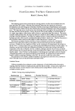
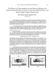
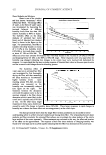
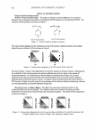
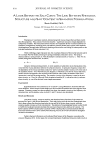
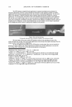
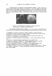
IMAGE ANALYSIS AND HAIR REMOVAL EFFICACY 347 addition to the use of the template, a side-by-side comparison of the first image taken at the following assessment times enabled optimized area relocation. AnalySIS® software (Soft Imaging System GmbH, Munster, Germany) was used to capture and process the images. Processing grey level images was done in order to discriminate visible hairs on the skin surface. To achieve this, a shading correction was applied to the grey images to reduce inhomogeneous background illumination. The images were filtered with optimized rank and sigma filters to reduce background noise and to resharpen the images. After discrimination by use of a dynamic threshold auto maticaUy adjusted to the different skin types, the parameters of hair length, hair width, and projection area were measured for each single hair in the images. The projection area gives a measure of the surface of the hairs, flattened by the measuring head. Hairs below a length of 200 µm were discarded in determination of all parameters to remove possible invalid data. In the case of hair length and hair width, overlapping hairs were not taken into account, while the projection area included overlapping hairs. RESULTS Figure 1 gives an impression of the data that can be derived from the high-resolution macrophotographs. Compared to the before-shaving state, hair stubble was already visible one day after shaving with a disposable blade (left images). After eight days, hairs were regrown to their original length. Wax depilation removes hairs deep in the follicle. However, some non-depilated hairs were still visible after depilation, as can be seen in Figure 1 (right images). Irritation is seen as a red spot around a non-depilated hair. After eight days, the irritation had disappeared. Figure 2 and Table I give an overview of the results for Group 1, comparing shaving to depilation over a period of nine days. In Figure 2a, the change in the number of hairs is presented. On day 2, one day after hair removal, the number of hairs clearly decreased on the shaved as well as on the depilated test sites, with a more marked decrease on the depilated site, as expected. In the following days, the number of hairs detected increased again, reaching baseline level on days 7 to 9 on the shaved sites. Figure 26 represents hair thickness, showing no marked changes from baseline level on the shaved as well as on the depilated areas, except for a slight increase on day 2. This increase can be attributed to a shift in relation between thick and thin hairs due to hair removal. Figures 2c and 2d represent the results of hair length and projection area. Both parameters show a comparable evolution: It was seen that hair length and projection area did not change markedly until day 9 on the wax-depilated test fields, while on the shaved areas the hair length and projection areas returned to the initial state by days 7 to 9. Regarding day 2, one day after hair removal, hair length did not show a clear difference between the shaved and depilated sites due to the removal of invalid data (see above, overlapping hairs). The area projection showed lower values on the depilated site compared to the shaved site. After wax depilation, a very slight increase in the depilated site in the projection area (Figure 2d) and in hair length (Figure 2c) was documented from days 7 to 9. A contributing factor to this increase could be the regrowth of incompletely depilated hairs. Such incomplete depilation was starting to become visible on days 2 and 4, one to three days after hair removal. According to the results shown in Figure 2, wax

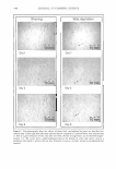
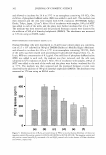
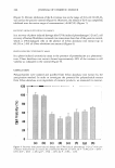
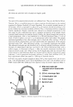
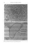
348 JOURi_ T .1. L 01� cos�rnTIC ... CTENCE Shaving Wax depilation 0. 0mm Day 1 Day 1 0 Day2 Day2 0mm Day9 Day9 Figure 1. :Macrophotographs from one subject of shaved and wax-depilated leg areas one day after hair removal (day 2) and eight days after hair removal (day 9) compared to condition before hair removal (day 1). Due to a good relocation of test areas after one week, the fate of single hairs can be observed (magni fication: 5 x). The circles show a small pigmented spot at the bottom of a hair that can be used as a permanent skin mark to relocate the test area. Irritation is seen as a red spot around a non-depilated hair (arro ·s).
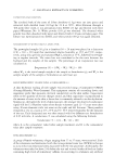
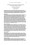
Purchased for the exclusive use of nofirst nolast (unknown)
From: SCC Media Library & Resource Center (library.scconline.org)