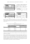
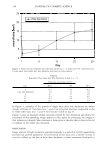
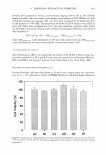
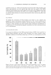
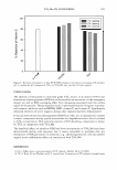
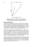
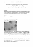
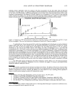
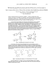
2006 ANNUAL SCIENTIFIC SEMINAR 411 The hysteresis factors for the forearm skin varied broadly from 17. 5% to 7 5%, with the highest value obtained for the oldest panelist. Excluding data obtained for the oldest panelist, the hysteresis factor of the skin of other subjects ranged from 17.5% to 38% and was similar in terms of viscoelastic contribution as compared to the skin models. A similar range of hysteresis factors ( or viscous coefficients), from 9. 3 % to 19% based on the analysis of three subjects, was reported by Zahuani et al. [2] In contrast to this, the strain energy data obtained for the periorbital region of the face suggest higher contribution of viscoelasticity with the hysteresis factor ranging from 35% to 49% . Conclusions: - The data from indentometric analysis of both artificial models of skin as well as forearm and periorbital skin of panelists obtained in-vivo can be interpreted by using Hertz theory of contact mechanics. The loading and unloading indentometric curves can be employed to calculate total and elastic strain energies as well as hysteresis ratio, which is the measure of viscoelasticity of an investigated material. - The indentometric creep and stress relaxation analysis of in-vivo skin and skin models can be described quantitatively by the Kelvin-Voigt model with one relaxation time. - The artificial skin models represent a good approximation of the behavior of in-vivo skin in terms of viscoelastic/elastic character. Skin models were, however, stiffer than natural skin with Young's modulae ranging from 5.5·104 N/m2 to 17.7·104 N/m2 for models vs. 0.7·104 N/m2 to 3.3 ·104 N/m2 for forearm and facial skin. In forthcoming studies we are planning to employ artificial skin models and natural skin to study the effect of thin polymer films on the mechanical behavior of skin. References [1] -KL.Johnson, Contact Mechanics, Cambridge University Press, 1985. [2] - H.Zahuani, C.Pailler-Mattei, R.Vargiolu, and M.A. Abellan, Assessment of the elasticity and tactile properties of the human skin surface by tribological tests, Proceedings of the 22nd IFSCC Congress in Edinburgh, 2002, podium 33.

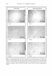
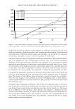
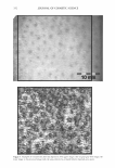
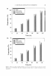
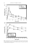
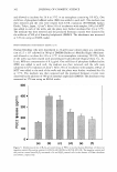
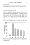
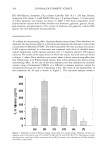
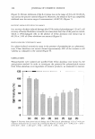
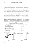
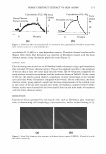
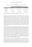
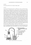
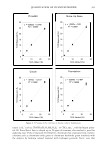
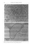
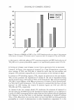
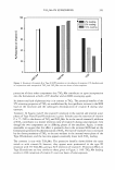
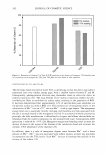
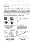
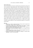
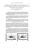
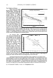
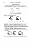
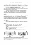
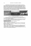
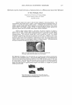
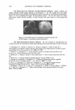
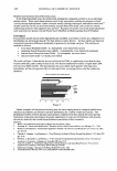
412 JOURNAL OF COSMETIC SCIENCE USE OF DIGITAL PHOTOGRAPHY AND IMAGE ANALYSIS TECHNIQUES TO ASSESS STRATUM CoRNEUM COMPROMISE IN HEALTH CARE WORKERS (HCW's) Jennifer Smith Canning1•2, Brian Barford3, Randy Wickett, Ph.D.2, and Marty Visscher, Ph.D. 1 1 The Skin Sciences Institute, Cincinnati, OH 2 University of Cincinnati, Cincinnati, OH 3 BCC Group, Cincinnati, OH Introduction Neonatal intensive care unit (NICU lp atienlli can experience complications due lo infection because of the risk of exposure lo invasive procedures and immature host defenoc mechanisms · . Hand hygiene is the most important measure of preventing healthcare-associated infections in critically ill neonates'· 5 , Standard hand h l giene guidelines are outlined by the Center for Diseaoc Control (CDC), but Compliance with the CDC hand hygiene gu idelines remains low, al about 30% . Skin irritation from repetitive hand hygiene procedures was reported as one of the main reasons for lack of compliance'. Frequent hand washing and repetitive exposure to soap/surfactant cleanocrs and water has si gn ificant effects on the structure and function of the stratum corneum (SC) barrier, e.g., drying, scaling, cracking and ocvere hand irritation 7, An increased IJSe in gloves and seasonal low humidity can also add lo the negative effeclli. Barrier damage pulli healthcare workers (HCW) at an increased risk of acquiring infection and therefore places patienls at an increased risk of nosocomial infections 7. The challenge is lo provide a regimen of products that maintain an effective SC barrier and facilitate hand hygiene compliance, Erythema (abnormal redness due lo dilation of capillaries) is often a sign of inflammation or infection. The degree of erythema ocen is often based upon subjective observations, i.e. Visual Skin Grading (VSG). The drawback is their low reliability and reproducibility. Skin erythema is difficult to assess in HCWs becauoc of their compromised SC. Therefore, there is a need for objective, quantitative techniques for the evaluation of skin color. The goal was to develop an accurate, cost-effective technique that could be used easily in our clinical setting. A digital imaging system was created and image analysis techniques were applied and the results compared with those from visual skin grading techniques. Methods HCWs from the Regional Center for Newborn Intensive Care (RICNIC) at Cincinnati Children's Hospital Medical Center (CCHMC) were the subjects in the Summer (n=54) and Winter (n=60)trials. Subjects had a history of skin damage, worked al least two consecutive 8-holU' shifts and performed al least 20 hand hygiene procedlU'Cs in an 8-holU' work shift. One of two treatment product sets was assigned at random. At the beginning and end of each of six work cycles per trial, high resolution digital images were taken of each hand and knuckles, fingers and dorsal surfaces were graded for erythema and dryne•s. lmages were also collected from control subjects with no visible skin damage and who did not undergo repealed hand hygiene procedures. A Fuji S2-Pro Camera, 6.1 Megapixels, SLR, a Nikon SB-29s Macro Speedlight flash and an AF Micro-Nikkor 60mm f/2.8 Macro Lens were used lo provide image resolution of 72 pixels/inch (2.8 pixels/mm). The hands were accurately repositioned lo allow comparison of images . All images were standardized for white balance and color with controlled lighting conditions. A subset of 25 subjects with the following criteria was used for image analysis: completed both trials, had noticeable contrasts oferythema between images, and had a wide range of visual grades (Range: 0.0-3.5 on a scale of 0.0-4.0). The right hand was chosen for image analysis (dominant for all subjects). Cycle 2 photos were uocd for image analysis since all subjects had used their designated products for about the same amount of time for both trials. Percentage of excess red pixels: lmages were optimized and separated into three channels (L, a• and b•, Adobe Photoshop). The a• image was used for further image analysis of erythema without the confounding effects of the full color. All pixels in the image of a• values were placed on a scale of O (least red) -255 (most red) based on redness intensity. A histogram of color was created from these values to produce a mean (11) and standard deviation(") of redness, along with a total number of pixels for each image (lmageJ). Pixels with redness higher than the 11+0- were used to represent excess redness in the image. The percentage of excess red pixels = (pixels higher than 11+0- /total # pixels)• 100%. Percent of excess red pixels was also calculated for 11, 11+20- and 11+30- for comparison. Area of Redness: The area of redness was calculated from the image histogram as another technique. This technique may be more reliable because of its familiarity (technique used previously in many other applications of science and mathematics.) I" moment calculation: ENi = sum of pixels higher than 11+0- 2 nd moment calculation: EiNi = sum of ((pixels higher than 11+0-) x (# on 0-255 scale (i))). Image Comparison: Two images were simultaneously compared (i.e. beginning versus the end of a work cycle) on a high resolution monitor (VPS software, BCC Group). Images were randomly presented with the control and test aides randomly switched without knowledge and with the images being presented in random order. The images were accessed by two expert skin graders. The VPS quantitative image comparisons were correlated with the other image analysis techniques and the visual skin grading. Results Using the live grading technique (VSG), the knuckles had significantly higher grades than the fingers and the dorsal area for both the summer and winter trials. Looking al VSG cycle differences in figure 3, knuckle erytherna increased during the work cycle in both trials and then decreased over the summer time off and increaocd further during the winter time off. Similar pattern• are seen for the fingers and dorsal area. The percent excess red pixel analysis and the visual perception (VPS) evaluation produced similar results. In figures I and 4, erythema decreased during the work cycle in the summer trial but increased duri ng the work cycle in the winter trial. Erythema increased in the winter aver time off while it decreased in the summer using VPS grading (increased slightly in the summer using percent excess red pixel analysis). Irritation was also examined using VPS along with erythema. Irritation results were very similar to the results in erythema, indicating a relationship between erythema and irritation. Visual skin grading results show some differences when compared with the other techniques. Winter VSG results are similar lo the other techniques, as occn in figure 3, but summer results show increased erythema during the work cycle and decreased erythema during time off whereas the other techniques tend to show decreased erythema during the work cycle and further decreaocd erytherna during time off in the summer. Though VSG does focus on differing areas of the hand while the other techniques focus on the hand as a whole, the other techniques may prove lo be more reliable. Visual skin grading is not a reproducible method while imaging methods can be reproduced and images can be taken much more efficiently. lmages can also be used in many different analyzing techniques and can be used lo find new analyzing techniques and new ways to look at problems that arioc with the skin.
Purchased for the exclusive use of nofirst nolast (unknown)
From: SCC Media Library & Resource Center (library.scconline.org)