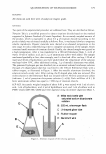
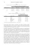
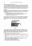
346 JOURNAL OF COSMETIC SCIENCE products to induce or improve hair growth increases. As a consequence, the development of methods to reliably quantify the efficacy of these products and methods is of increas ing interest. In classical dermatology, clinical scoring (2 ,3) and trichogram analysis by the plucking of hairs (4-6) are the methods usually used to assess hair growth patterns on the human scalp. They are suitable when the focus is on diagnosis of scalp hair diseases like alopecia. For assessment of depilation efficacy or hair growth inhibition, advanced imaging tech niques are more suitable and can be used on all body sites of interest, such as legs, axilla, and face. Standardized clinical photography and image analysis to assess hair growth in a non invasive way has been used since 1970 (7) and has improved continuously (8-12). To date, the computer-aided capturing of high-resolution images offers direct control of magnification, image section, and image quality. Modern, powerful, image-analysis software packages (13) enable the user to program tailor-made parameters for specific needs. Retrieval of single hairs in images and the repeated measurement of their length, width, and projection area are powerful tools to quantify hair growth. The closeness of a depilation method's giving a measure of the method's efficacy, the time until hairs become again visible after their removal, growth velocity, and even skin irritation can be quantified. Taking wet shaving and wax depilation as well known examples, we dem onstrate how depilation techniques or actives designed to reduce hair growth can be benchmarked with the help of improved image analysis. MATERIALS AND METHODS In two test panels, Group 1 consisting of ten and Group 2 consisting of nine female volunteers, between 25 and 65 years of age, hair removal methods were applied and investigated. As a pretreatment, the volunteers shaved their legs with disposable blades under the supervision of a technician seven days before starting the study, as a stan dardized starting point. On day 1, test areas of 3 cm x 3 cm were outlined on the inner sides of the lower legs, close to the tibia. Group 1 shaved one randomly assigned test area (right or left leg) the other leg was depilated with a cold wax (marketed product). In Group 2, only one test area was outlined on the lower leg (either right or left leg according to a randomization scheme). On day 1, the test area was depilated by using cold wax. In Group 1, images of the test areas were taken on study day 1 before hair removal, and on study days 2, 4, 7, and 9. In Group 2, images were taken on study day 1 before hair removal, directly after depilation, and then weekly over a period of four weeks. Macrophotographs (magnification: 5X) were taken with a high-performance stereomi croscope (Olympus SZX Series, Hamburg, Germany) equipped with a high-resolution CCD color camera (SIS Color View CC-12, 1.4 megapixel). An Olympus ring light connected to the objective tube (Olympus SZX Series, Hamburg, Germany) was used to enable homogeneous illumination. To be able to assess the same test area at all assessment times with an accuracy of better than 2 millimeters, a transparent template was prepared using permanent skin marks such as nevi, in or near the test area, as demarcation points. The template was a commercially available foil made of polyethylene with a thickness of 0.08 mm. In

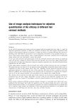
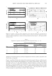
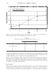
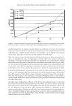
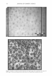
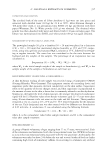
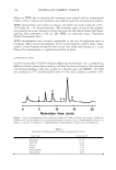
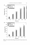
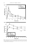
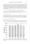
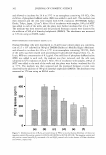
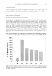
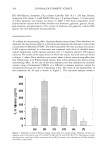
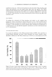
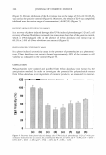
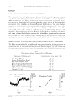
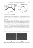
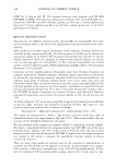
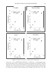
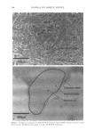
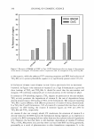
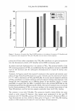
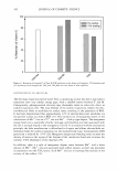
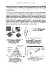
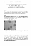
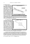
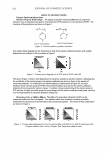
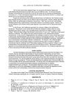
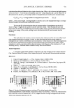
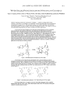
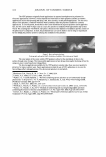
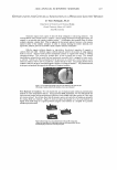
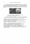
IMAGE ANALYSIS AND HAIR REMOVAL EFFICACY 347 addition to the use of the template, a side-by-side comparison of the first image taken at the following assessment times enabled optimized area relocation. AnalySIS® software (Soft Imaging System GmbH, Munster, Germany) was used to capture and process the images. Processing grey level images was done in order to discriminate visible hairs on the skin surface. To achieve this, a shading correction was applied to the grey images to reduce inhomogeneous background illumination. The images were filtered with optimized rank and sigma filters to reduce background noise and to resharpen the images. After discrimination by use of a dynamic threshold auto maticaUy adjusted to the different skin types, the parameters of hair length, hair width, and projection area were measured for each single hair in the images. The projection area gives a measure of the surface of the hairs, flattened by the measuring head. Hairs below a length of 200 µm were discarded in determination of all parameters to remove possible invalid data. In the case of hair length and hair width, overlapping hairs were not taken into account, while the projection area included overlapping hairs. RESULTS Figure 1 gives an impression of the data that can be derived from the high-resolution macrophotographs. Compared to the before-shaving state, hair stubble was already visible one day after shaving with a disposable blade (left images). After eight days, hairs were regrown to their original length. Wax depilation removes hairs deep in the follicle. However, some non-depilated hairs were still visible after depilation, as can be seen in Figure 1 (right images). Irritation is seen as a red spot around a non-depilated hair. After eight days, the irritation had disappeared. Figure 2 and Table I give an overview of the results for Group 1, comparing shaving to depilation over a period of nine days. In Figure 2a, the change in the number of hairs is presented. On day 2, one day after hair removal, the number of hairs clearly decreased on the shaved as well as on the depilated test sites, with a more marked decrease on the depilated site, as expected. In the following days, the number of hairs detected increased again, reaching baseline level on days 7 to 9 on the shaved sites. Figure 26 represents hair thickness, showing no marked changes from baseline level on the shaved as well as on the depilated areas, except for a slight increase on day 2. This increase can be attributed to a shift in relation between thick and thin hairs due to hair removal. Figures 2c and 2d represent the results of hair length and projection area. Both parameters show a comparable evolution: It was seen that hair length and projection area did not change markedly until day 9 on the wax-depilated test fields, while on the shaved areas the hair length and projection areas returned to the initial state by days 7 to 9. Regarding day 2, one day after hair removal, hair length did not show a clear difference between the shaved and depilated sites due to the removal of invalid data (see above, overlapping hairs). The area projection showed lower values on the depilated site compared to the shaved site. After wax depilation, a very slight increase in the depilated site in the projection area (Figure 2d) and in hair length (Figure 2c) was documented from days 7 to 9. A contributing factor to this increase could be the regrowth of incompletely depilated hairs. Such incomplete depilation was starting to become visible on days 2 and 4, one to three days after hair removal. According to the results shown in Figure 2, wax
Purchased for the exclusive use of nofirst nolast (unknown)
From: SCC Media Library & Resource Center (library.scconline.org)