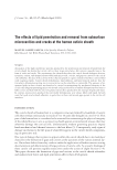
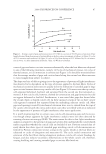
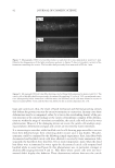
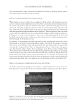
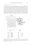
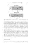
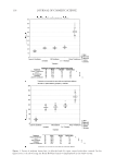
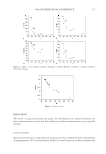
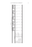
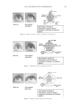
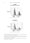
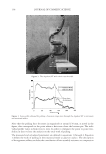
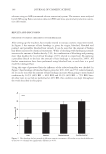
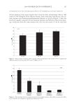
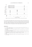
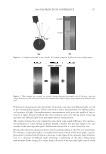
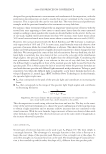
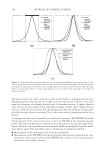
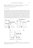
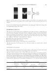
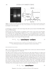
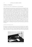
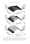
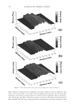
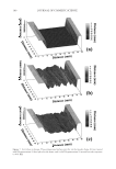
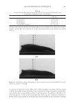
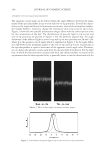
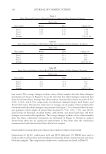
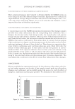
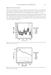
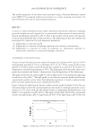
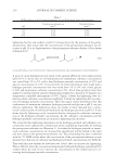
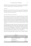
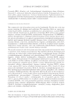
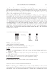
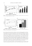
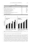
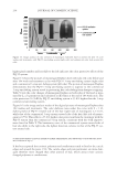
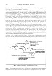
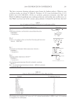
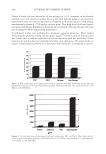
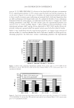
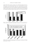
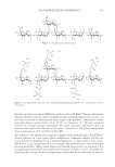
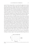
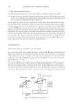
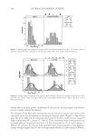
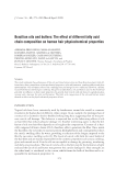
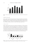
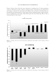
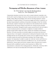
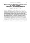
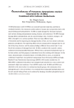
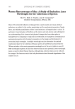
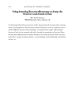
2008 TRI/PRINCETON CONFERENCE 203 Figure 5. Comparison of spectra from sample parallel and perpendicular to plane of polarization. Middle spectrum: long axis of fi ber parallel to plane of polarized laser. Bottom spectrum: long axis perpendicular to polarization. Top spectrum: Difference spectrum (parallel – perpendicular). In summary, this work demonstrates that 780 nm is the most appropriate excitation wavelength to use to investigate hair samples by Raman spectroscopy. The spectra collected when the fi ber is oriented parallel and when oriented perpendicular to the direction of polarization of the excitation laser are shown in Figure 5: the difference spectrum of the two is also shown. The Raman spectra recorded are observed to be sensi- tive to the orientation of the hair sample with respect to the laser polarization. The difference spectra features two a-helical at 1655 cm−1 (Amide I) and 935 cm−1 (C-C stretch), together with the Cα-H bending mode at 1314 cm−1 as being the most orienta- tion sensitive bands. As the anisotropic component of the fi ber is comprised mainly of α-helical intermediate fi laments, these results suggest that the relative intensities at 1655 cm−1 and 935 cm−1 could be used as a measure of alignment of the α-helical intermediate fi laments with the fi ber axis. This could be exploited to explore structural differences between hair samples of different ethnicity or hair subjected to different treatments. CONCLUSION The multiple wavelength experiments show that the 780-nm excitation is the most ap- propriate wavelength out of the set of visible-to-NIR wavelengths explored. It has been

JOURNAL OF COSMETIC SCIENCE 204 observed that while shorter wavelength lasers can be used to obtain greater signal inten- sity, potentially reducing collection times, the shorter wavelength causes a large increase in background intensity, negating this benefi t. Exploratory experiments designed to determine the applicability of polarized Raman show changes in the spectra depending on orientation of the fi bers to the direction of the exciting beam. This may therefore offer potential to explore and estimate orientation of the crystalline intermediate fi laments in hair fi bers. REFERENCES (1) C. M. Pande. FT-Raman spectroscopy—Applications in hair research, J. Soc. Cosmet. Chem., 45, 257–268 (1994). (2) F. J. Wortmann, C. Popescu, and G. Sendelbach. Effects of reduction on the denaturation Kinetics of human hair, Biopolymers, 89, 600–605 (2008). (3) J. L. Haston, S. B. Engrlsen, M. Roessele, J. Clarkson, E. W. Blanch, C. Baldock, C. M. Kielty, and T. J. West, Raman microscopy and X-ray diffraction, a combined study of fi brillin-rich microfi brillar elasticity, J. Biol. Chem., 278(42), 41189–41197 (2003). (4) A. Kuzuhara, Analysis of structural changes in keratin fi bers resulting from chemical treatments using Raman spectroscopy, Biopolymers, 77, 335–344 (2005). (5) K. Schaefer. Natural fl uorescence of wool, J. Soc. Dyers Colours, 107(5–6), 206–211 (1991). (6) K. Song and J. F. Rabolt, Polarized Raman measurements of uniaxially orientated poly(ε-caprolactam), Macromolecules, 34, 1650–1654.
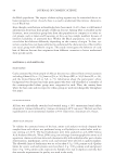
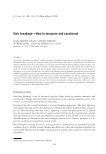
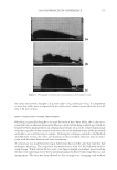
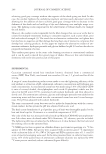
Purchased for the exclusive use of nofirst nolast (unknown)
From: SCC Media Library & Resource Center (library.scconline.org)