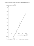
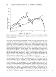
J. Soc. Cosmet, Chem., 29, 177-184 (March 1978) Measurement of enzyme kinetics on the intact skin a new method to study the biological effects of cosmetics on the epidermis PETERT. PUGLIESE Xienta, Inc., R.D. #1, Bernville, PA 19506. Received December 7, 1977. Presented at 9th International Congress, IFSC C,June 1976, Boston, Mass. Synopsis A new DIRECT FLUOROMETRIC method allows the MEASUREMENT of ENZYME ACTIVITY on the INTACT SKIN of various body surfaces. This permits normal physiological parameters to operate on the system studied. The pentose phosphate pathway (Entner-Doudoroff), previously shown to operate in the epidermis, provides several enzymes which are used here to assess and compare various dermatological con- ditions. Glucose-6-phosphate dehydrogenase and lactic dehydrogenase are measured by fluorometric termination of changes in NADPH and NADH. Other enzymes of the Embden Myerhoff and Krebs cycles are measured directly or indirectly by this method. Various COSMETIC base ingredients and compounded formulations were studied to determine their EFFECTS on epidermal metabolism. Enzyme action was recorded as increased, decreased or not affected. This new method is simple and relatively inexpensive, and allows extremely wide applications. INTRODUCTION The need for new methods to study the biochemistry of the epidermis in situ is becom- ing more apparent. Methods involving extrapolation from animal models are not al- ways applicable to the human epidermis because of species differences. Excised skin from human volunteers has certain inherent drawbacks including pain, disfigurement and the problem of an isolated specimen removed from its normal milieu. The use of an in vivo and in situ method would obviate many of these problems. This paper describes such a method. The skin is the largest body organ, representing approximately one-sixth of the body weight. Far from being a mere barrier to the internal and external environment, the skin is proving to be a dynamic organ with a profound effect on the internal me- tabolism. A review of the extensive literature on the metabolic functions of the epi- dermis reveals carbohydrate metabolism to be unique (1-4). For this reason we chose epidermal carbohydrate metabolism as our biochemical system. Previous studies have demonstrated enzyme activity in histochemical sections (5, 6), cell homogenates (7) and epidermal stripping (8). These experiments have been both comprehensive and 177

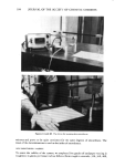
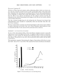
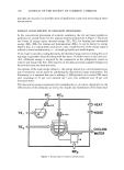
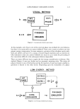
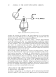
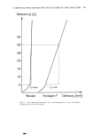
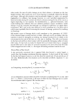
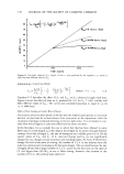
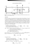
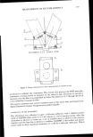
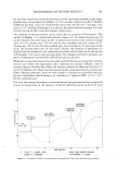
178 JOURNAL OF THE SOCIETY OF COSMETIC CHEMISTS elegant with conclusive demonstration of enzyme activity in the upper layers of the epidermis. Recently Schalla et•/. (9) demonstrated enzyme activity in the intact epi- dermis by perfusion of a glass chamber attached to the surface of the skin in situ. Their measurements were recorded as a change in absorbance of the perfusate in a flow- through spectrophotometer. Early studies by Chance et M. (10) have shown that fluorescence changes can be de- tected in the intact organ and that these changes reflect a state of oxidation-reduction within the organ cells. This study and prior studies by Chance et M. (11) indicate that tissue irradiated at 366 • emitted fluorescence characteristic of the reduced pyridine nucleotides. The preponderance of nucleotide fluorescence in the tissues precludes the measurement of other cellular fiuorophores with similar fluorescence characteristics. It was further concluded that the assay of reduced pyridine nucleotides is insensitive to the state of oxygenation of hemoglobin and thus this system can be used to follow oxi- dation-reduction ratios in anoxic states. Based on these previous studies, we designed a system that would measure fluorescence changes of the pyridine nucleotides involved in carbohydrate metabolism in the epidermis. METHODS AND MATERIALS THE FLUOROMETER Our system employs a sensitive fluorometer that measures the change in fluorescence of nicotinamide adenine dinucleotide (NAD) or nicotinamide adenine dinucleotide phosphate (NADP) when they are in the reduced state, NADH or NADPH, respec- tively. Excitation is at 340 to 365 nm and emission is at 460 nm. In oxidized form (NAD or NAPD) they are not fluorescent. The emission intensity is directly propor- tional to the amount of reduced nucleotide. Measurements of changes in fluorescence, either increased or decreased, can be utilized with this system. Our instrument em- ployed the electronic components of the Metabolite Fluorometer E704, designed and built at the Johnson Foundation of the University of Pennsylvania. Essentially, this instrument employs a GEF4T4 Germicidal UV lamp with a E062 socket. The ul- traviolet light from this source is filtered through a Corning No. 5840 filter or Wratten 18A Kodak filter. The visible emitted light is detected by a photomultiplier tube (PMT) EM19524B (supplied by EMI Gencom Inc., 80 Express Street, Plainview, New York). Ultraviolet light is filtered out by inserting a UV filter 2B (Kodak) or a Corning No. 3389 filter at the face of the PMT. Power supply to the PMT is provided by a Model 6515A DC Power Supply from Hewlett Packard. The recorder is a Honeywell Electronik 19 with 100 mv sensitivity, usually set at a speed of 0.5 in./min. The amplifier and output circuit diagram for the E704 is available from Johnson Founda- tion, University of Pennsylvania, Philadelphia, Pennsylvania. For our instrument, we designed the metabolite chamber and detection systems to allow direct measurement on the skin as follows (Figure 1). An aluminum block (D) was machined to allow insertion of the ultraviolet light source (E) and photomultiplier (F) with respective filters (G and H) at an angle of 22.5 ø from the perpendicular. The bottom of the block was machined to accommodate a closure mechanism (B) and a reaction chamber (A). The slide closure mechanism (C) and the reaction chamber are of stainless steel. By closing the reaction chamber, in •:itro enzymatic reactions can be
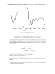
Purchased for the exclusive use of nofirst nolast (unknown)
From: SCC Media Library & Resource Center (library.scconline.org)