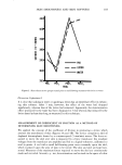
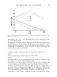
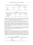
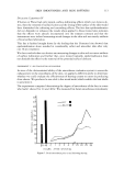
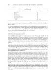
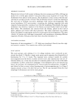
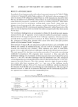
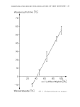
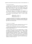
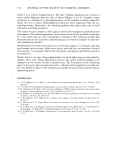
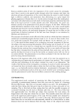
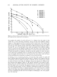
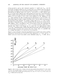
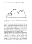
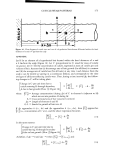
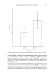
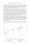
MEASUREMENT OF ENZYME KINETICS 183 the pyridine nucleotides in the first few layers of the epidermis, probably in the upper granular layer. It was shown by Schalla eta/. (9) in studies carried out with C 14 labelled NAD that less than 1 per cent of the NAD crossed the cell wall (10 -11 mol/mg), com- pared to 10 -7 mol/mg of hydrogen. It is obvious therefore that the hydrogen ion is the actively moving ion that causes the change in fluorescence. The question of bacteria activity can be raised also as a source of fluorescence. The studies of Marpies (13) on skin flora indicate a figure of 1.05 million bacteria per m" on the forearm. Our own values on the 12 subjects used in the study yield an average value of 10.6 colonies over 120 cm.' area for the unwashed arm and a value of 1.1 colonies per 120 cm" for the water-washed arm. Since less than 4 cm.' were used in the study, the bacteria effect may be discounted. Finally, the question of absorbancy of liquids from the chamber by the epidermis may fairly well be ruled out by the studies of Scheuplein (14) on percutaneous absorption. The changes observed in this study are almost instantaneous and precluded diffusional rates mechanisms. While the enzyme rates reported in this study are far below those reported for cell-free extracts and whole-cell suspensions, they represent an entirely different class of enzyme kinetics. Possibly they follow the kinetics outlined by Blum and Jender (15) and by O'Sullivan (16) which deal with geometrically constrained enzyme systems and slowly diffusing substrates. Since we were unable to calculate an actual Krn value for glucose-6-phosphate dehydrogenase, we calculated an "apparent KM" of 2.9 x 10 -4 for the conditions used. One very interesting observation on several patients with psoriasis was the unexpected /ncrease in fluorescence in the presence of partial epidermal anoxia induced by local 8C •60 ADD G-6-P ll.6 q MOLES SKIN TOURNIQUET RELEASED ß TOURNIQUET /' APPLIED '"' , ADD NADP I " ?_5 mq MOLES ,,' ß TIME• MINUTES SUBJECT A (NORMAL SKIN) SUBJECT B (PSORIASIS) RIGHT ARM, DORSAL SURFACE POSTPRANDIAL- 2 HOURS Figure 3. Comparison of effect of partial anoxia in normal individuals and individuals with psoriasis

184 JOURNAL OF THE SOCIETY OF COSMETIC CHEMISTS venous occlusion. Partial anoxia causes a decrease in fluorescence in the normal indi- vidual. This is seerl in Figure 3. At present, we have no explanation for this phenomenom. It is found only in patients with• active psoriasis and quickly returns to normal when the skin is treated with steroids, or exposed to sunlight for 4 to 6 hr. The ef- fect appears to be related to the severity of the disease, though we have observed itin indi- viduals who have inactive psoriasis and are not under treatment of any type. A- great deal of additional work needs to be done on this system before it will become a routine procedure. The ease of performing the test, the lack of epidermal invasion, and the use of human subjects should make the procedure generally applicable for the study of epidermal effects of applied agents. CONCLUSIONS 1. A new in vivo and in situ method for studying epidermal biochemistry has been out- lined. 2. The method is reproducible and correlates with similar methods--that is•perfu- sion and stripping techniques. 3. Classical Michaels-Menten kinetics do not appear to hold for this technique. Precise kinetic studies are needed. 4. Applications appear almost limitless, since the epidermis is accessible and the method adaptable to any reaction in which fluorescence or absorption changes can be measured. REFERENCES (1) D.M. Pillsbury, The intrinsic carbohydrate metabolism of the skin, J. Amer. Med. Ass., 96,426(1931). (2) E. Urbach and J. W. Lentry, Carbohydrate metabolism of the skin, Arch. Dermatol. Syphilol., 52, 301(1945). (3) A. Jarrett, The penrose phosphate pathway in human and animal skin, Brit. J. Dermatol. Syph., 84, 545(1971). (4) P. D. Mier and D. W. Cotton, "The Molecular Biology of Skin," Blackwell Scientific Publication, Ox- ford, England, 1976, Chapter 2. (5) F. B. Hershey, Quantitative histochemistry of skin, J. Histochem. Cytochem., 8, 41(1960). (6) K. Adachi and S. Yamasawa, Quantitative histochemistry of pyruvate skin, J. Invest. Dermatol., 46, 473(1966). (7) R. K. Freinkel, Metabolism of glucose-C-14 by human skin in vitro, J. Invest. Dermatol., 34, 37(1960). (8) M. Kermici, C. Bodereau and G. Aubin, Measurement of biochemical parameters in the stratum cor- neum, J. Soc. Cosmet. Chem., 28, 151(1977). (9) W. Schalla, A. Zesch and H. Schaefer, The estimation of enzyme activity in living epidermal cells, Brit. J. Dermatol. Syph., 91, 84a(1974). (10) B. Chance and B. Schoener, A correlation of absorption and fluorescence changes in ischemia of the rat liver, in vivo,Biochem. Zh., 341,340(1965). (11) B. Chance, P. Cohen, F. Jobsis and B. Schoener, Science, 137,499(1962). (12) O. H. Lowry, N. R. Roberts andJ. I. Kappan, The fluorometric measurement ofpyridine nucleotide, J. Biol. Chem., 224, 1047(1957). (13) M.J. Marpies, Life on the human skin,Sci. Amer., 220(1), 108(1969). (14) R.J. Scheuplein, Mechanism of percutaneous absorption, J. Invest. Dermatol., 48(1), 19(1967). (15) J. J. Blum and D. J. Jender, Rate behavior and concentration profiles in geometrically constrained enzyme system, Arch. Biochem. Biophys., 66, 316(1957). (16) D. G. O'Sullivan, Quantitative potentials of enzyme cytochemistry modified Michaels-Menten rate law applicable when a substrate diffuses slowly into an enzyme site, J. Theor. Biol., 2, 117(1962).
Purchased for the exclusive use of nofirst nolast (unknown)
From: SCC Media Library & Resource Center (library.scconline.org)