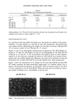
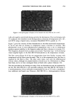
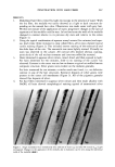
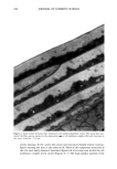
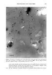
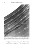
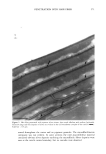
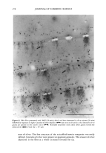
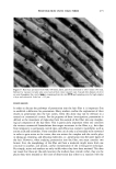
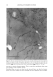
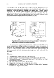
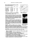
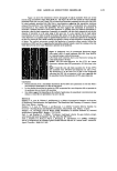
PENETRATION INTO HAIR FIBER 279 macrofibrillar material. What is very evident from these studies is that the medulla, at the center of the hair, the pigment granule within the body of the cortical cell, and the microfibril/matrix composite can all be accessed within one to two minutes or less at room temperature. It is also clear from these studies that the previous debate concerning the mode and route of penetration into the hair fiber needs further and careftA consideration. Much of the discussion is limited to considering the cuticle as a boundary structure that needs to be crossed. While this is true, it should also be viewed as a highly important compartment in its own right. Understanding the uptake and retention of materials into this com- partment and the transport of materials, via the cmc, across the cuticle depends on how the observations are made. It is most probable that access to the cortex is via the cuticle cell membrane complex. This multilamellar structure would favor transport parallel to the lamellae rather than normal to the lamellae. The suggestions by other authors that transport to the cortex occurs from the endocuticle would require molecules to traverse the cmc normal to its complex physicochemical lameliar structure. As yet we have no evidence for this route. CONCLUSION These studies have shown that aqueous molecules can be observed in all of the structures of the hair from one to two minutes after immersion in water-soluble reagents. Impor- tantly, these can be identified within the cuticle cell membrane complex and the bulk of the cuticle cell at the same time, indicating different but complementary pathways into the hair fiber. No evidence was found to suggest a diffusion pathway from the bulk of the cuticle cell, across the cmc, and into the cortex. Conversely, evidence was found for pathways from the cmc into the bulk of the cuticle cell along the whole of the hair fiber. By combining the correct reagents, a detailed knowledge of the hair structure, knowl- edge of the results and problems of product formulations, and, importantly, typical TEM stain results, one can build a more comprehensive view of penetration into the hair fiber. It is imperative to understand that the observation of a material in a particular structure does not always tell you how it got there. Nor does its absence from other structures say that it was never there. Importantly, penetration should be viewed as the movement of molecules from the outside of the fiber to various compartments in the fiber, rather than just from the outside of the hair to the inside of a cortical cell. ACKNOWLEDGMENTS I gratefully acknowledge Miss Patricia Goggin, Royal Holloway and Bedford College, for her technical assistance. REFERENCES (1) J. A. Swift, "The Detection of Pores and Holes in Hair by Electron Microscopy," in Hair Research for the Next Mi//eni•m, D.J.J. Van Neste and V. A. Randall, Eds. (Elsevier Science BV, Amsterdam, 1996), pp. 109-112.

280 JOURNAL OF COSMETIC SCIENCE (2) J. A. Swift, Further comments on "Pathways for aqueous diffusion in keratin fibres," Text. Res. J., 70, 277-278 (2000). (3) F.-J. Wortmann, G. Wortmann, and H. Zahn, Pathways for dye diffusion in wool fibres, Text. Re•. J., 67, 720-724 (1977). (4) J. D. Leeder, J. A. Rippon, F. E. Rothery, and I. W. Stapleton, Use of transmission electron microscope to study dyeing and diffusion processes, Proc. 7th Int. Wool Text. Res. Conf, Tokyo, 5, 99-108 (1985). (5) J. D. Leeder, Comments on "Pathways for aqueous diffusion in keratin fibres," Text. Res. J., 69, 229 (1999). (6) J. A. Swift, Electron histochemistry ofcystine containing proteins in thin transverse sections of human hair. J. Royal Microscop. Soc., 88, 449 (1968). (7) C.L. Gummer, R. P. R. Dawber, and V. H. Price, Trichothiodystrophy: An electron-histochemical study of the hair shaft, Br. J. Dermatol., 110, 439-449 (1984).
Purchased for the exclusive use of nofirst nolast (unknown)
From: SCC Media Library & Resource Center (library.scconline.org)