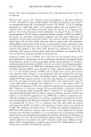
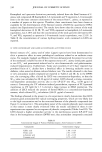
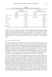
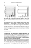
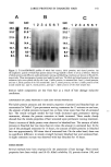
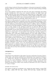
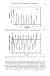
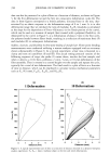
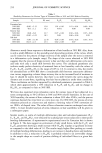
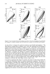
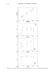
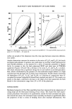
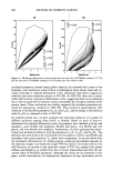
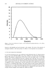
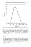
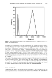
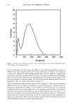
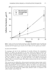
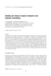
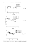
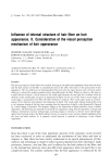
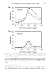
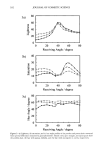
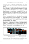
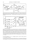
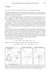
390 JOURNAL OF COSMETIC SCIENCE shouldered single peak and diffuse scattering in a wide range of receiving angles. The main peak is the specular reflection from the front surface, and the second peak or the shoulder is the specular reflection from the back surface. The two reflections from the front and back surfaces are separated by inclined cuticle surfaces (6-9). The front surface reflection is affected only by the optical properties of the hair surface, such as surface roughness. On the other hand, the back surface reflection is affected by the optical properties inside the hair fiber, adding to the surface properties. The back surface reflection is refracted at the front surface, reflected at the back surface, scattered at the porous structure when it exists, and also absorbed by melanin granules and/or dyestuffs when they exist. Figure 2 shows the gonio profiles at 700 nm and 400 nm of the hair samples charac- terized in Figure 1: poreless hair (closed circle), the hair with porous medulla (open triangle), and the hair with micropores in cortex (open square). The wavelength at 700 nm (Figure 2a) was selected to get information on the internal structures, because the light penetrated into the fiber is less absorbed by melanin granules at a longer wave- length than at a shorter wavelength (10). On the other hand, the wavelength at 400 nm (Figure 2b) was selected to get information mainly on the surface structure, because the influence of the internal structure is minimized depending on the high absorption ability of melanin granules at a shorter wavelength. The differences in the goniophotometric profile at 700 nm shown in Figure 2a are expressed in terms of light scattering inside the hair fiber. The diffuse scattering is clearly observed in the region from 0 to 30 degrees of the receiving angles, while the back surface reflection appears to be at 40 to 80 degrees and overlaps on the front surface reflection at around 40 degrees. Therefore, the apparent peak intensity of the front surface reflection of each hair looks different. In the case of the poreless hair, the light penetrating into the hair fiber is scattered less, leading to low levels of diffuse scattering and a strong back surface reflection, in comparison to the porous hairs. The peak intensities of the goniophotometric profiles at 400 nm in Figure 2b show little difference between the poreless hair (closed circle) and the hair with porous medulla (open triangle), meaning that the surface roughness is not different significantly between the two types of hair. The hair with micropores in the cortex (open square) gives a smaller and broader peak compared to the other hairs, because the surface of this hair is rougher than the above-mentioned two types of hair. In order to understand visual brightness and color perceptions, spectral reflectance (390-700 nm) at each angle is converted to lightness, saturation, and hue angle in the CIE color system (Figure 3). Each profile of lightness (Figure 3a) shows a single peak at 40 degrees, the front surface reflection in Figure 2. On the other hand, the saturation profiles (Figure 3b) show a peak around 60 degrees, corresponding to the back surface reflection. In the case of the poreless hair (closed circle), higher contrasts in the lightness and saturation are observed. The hairs with porous medulla (open triangle) and with micropores in cortex (open square) show broader peaks of lightness and saturation than the poreless hair, meaning a lower contrast. The two kinds of contrast in the lightness and the saturation are in the following order: poreless porous medulla pores in cortex, in the present case. The data of the hue angle (Figure 3c) also show a similar effect in the porous structure. In the case of the porous hairs (open triangle and square), the profiles of the hue angle

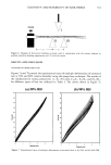
HAIR APPEARANCE AND INTERNAL STRUCTURE 391 2a) 0.5 0.4 0.3 0.2 0.1 0.0 700 nm ...... o ..... [] ,• -"o...'...•_ .... o ..... o ..... '-' ,'½'-2' '•'""'o,,.... i i i i i I i 0 20 40 60 80 Receiving Angle / degree 2b) 0.5 0.4 0.3 400 nm 0.2 0.1 0.0 0 20 40 60 80 Receiving Angle / degree Figure 2. Goniophotograms for poreless and porous hairs at (a) 700 nm and (b) 400 nm wavelength. Closed circle, open triangle, and open square show the poreless hair, the hair with porous medulla, and the hair with micropores in cortex, respectively. are not changed so much with the receiving angle. Only the hue angle of the poreless hair (closed circle) shows a bigger difference in the receiving angles from 0 to 40 degrees. This change in hue angle means that the apparent color is changed with the observing angle. VISUAL APPEARANCE OF HAIR SAMPLES The visual appearances of the poreless hair, the hair with a porous medulla, and the hair with micropores in cortex in Figure 1 were evaluated by professional panels, in order to
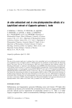
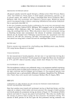
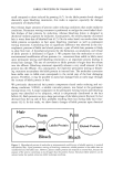
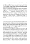
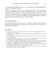
Purchased for the exclusive use of nofirst nolast (unknown)
From: SCC Media Library & Resource Center (library.scconline.org)