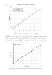
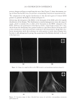
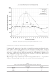
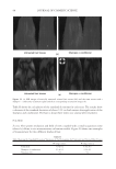
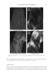
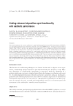
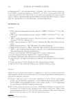
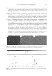
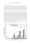
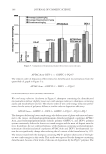
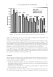
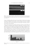
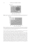
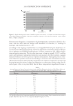
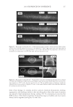
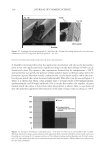
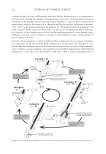
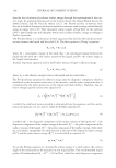
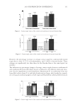
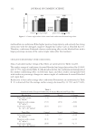
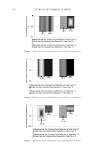
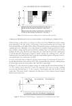
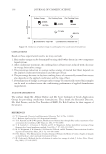
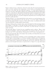
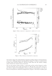
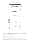
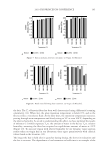
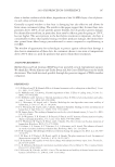
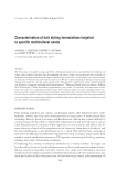
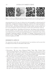
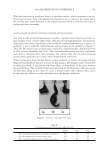
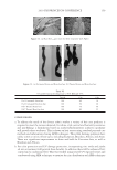
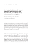
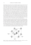
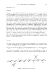
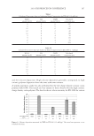
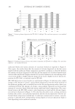
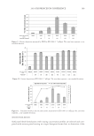
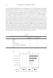
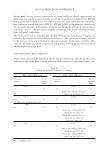
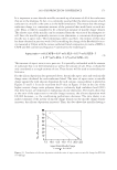
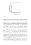
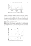
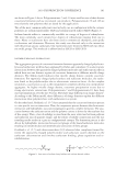
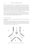
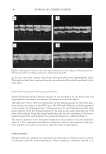
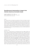
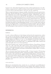
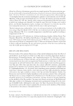
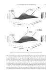
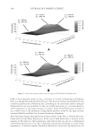
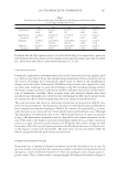
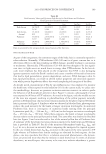
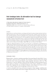
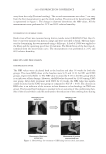
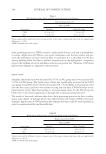
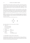
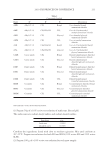
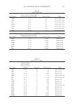
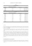
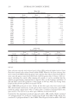
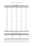
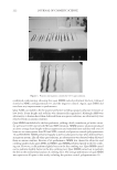
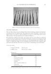
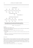
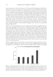
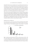
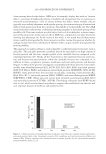
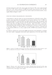
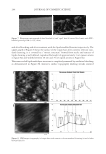
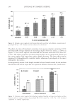
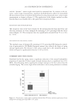
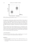
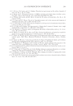
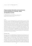
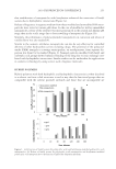
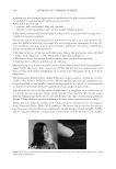
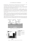
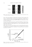
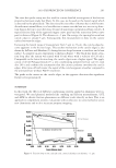
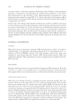
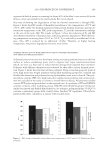
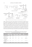
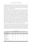
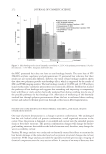
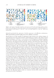
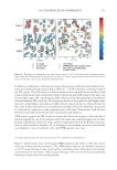
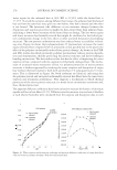
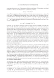
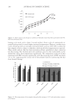
2010 TRI/PRINCETON CONFERENCE 243 the lipid composition and in the contact angle were detected. The contact angle dropped from 106° to 68° and the content of 18-MEA decreased by approximately 24%. The ultra bleaching process removed signifi cantly more surface lipids and showed smaller contact angles shown in Figures 5 and 6. DETECTION OF SURFACE CHANGES RELATED TO PROTEOLIPIDS A high affi nity to the hair surface for the proteolipid SR was confi rmed by means of fl uo- rescence microscopy. Hair was immerged into an aqueous solution of 0.15 % FITC-la- belled proteolipid for 5 min and rinsed out under tap water for 3 min. An intensive and widespread surface coverage was detected for the ultra bleached hair (Figure 7a). Areas of cuticle edges show the highest fl uorescence intensity. “Aged” hair shows a much more inhomogeneous light distribution. Here the fl uorescence intensity is mainly located at cuticle edges (Figure 7b). These differences in the fl uorescence intensity correspond well to the obtained differences in contact angles and the 18-MEA losses reported in Figures 5 and 6. In addition to fl uorescence microscopy SFM investigations were performed to visualize the cuticle surface topography and possible lipid organization at the hair surface before Figure 5. Amount of 18-MEA extracted from hair before (untreated) and after aging simulation (“aged hair”) in comparison to the amount of 18-MEA extracted after ultra bleaching (ultra bleached). Figure 6. Dynamic contact angles determined according to Wilhelmy before (untreated) and after aging simulation (“aged hair”) in comparison to the contact angle of ultra bleached hair (ultra bleached) (**p 0.01 calculated between “before aging” and the samples).

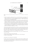
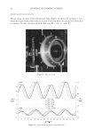
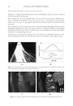
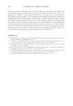
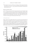
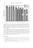
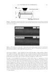
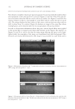
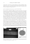
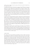
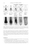
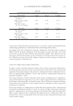
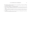
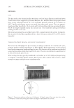
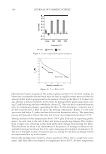
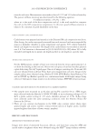
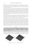
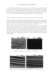
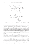
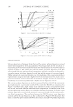
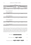
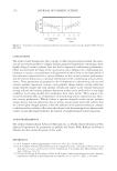
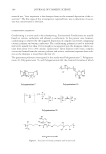
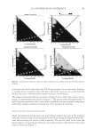
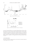
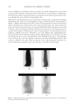
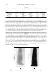
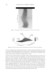
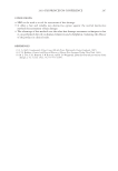
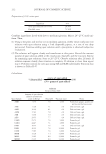
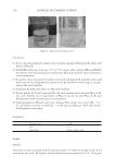
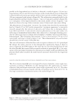
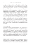
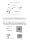
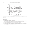
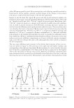
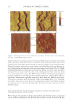
JOURNAL OF COSMETIC SCIENCE 244 and after bleaching and after treatment with the lipid-modifi ed keratins respectively. The upper graph of Figure 8 shows the surface of the virgin hair after cosmetic relevant stan- dard cleansing it is covered by a “mosaic structure” formed from stacks and terraces of lipids showing a well-defi ned, reproducible height of approximately 3 nm (green arrows in fi gure 8a) and widths between 20 nm and 70 nm (pink arrows in Figure 8a). This nano-scaled lipid multilayer structure is completely removed by oxidative bleaching as demonstrated in Figure 8b. Instead a surface topography showing streaks oriented Figure 7. Fluorescence micrograph of ultra bleached (a) and “aged” hair (b) treated (for 5 min) with FITC- labelled proteolipid SR (0.15%, m/m). Figure 8. SFM images (topography) of virgin hair with cosmetic relevant standard cleansing (a) and of ultra bleached hair (b).
Purchased for the exclusive use of nofirst nolast (unknown)
From: SCC Media Library & Resource Center (library.scconline.org)