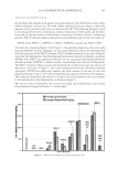
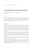
J. Cosmet. Sci., 62, 109–120 (March/April 2011) 109 Void and pore formation inside the hair cortex by a denaturation and super-contraction process occurring during hair setting with hot irons MANUEL GAMEZ-GARCIA, BASF Care Chemicals, 500 White Plains Rd, Tarrytown, NY 10591. Synopsis An analysis of hair fi bers from donors that frequently use hot irons for hair straightening showed the presence of multiple pores and voids (φ ∼0.1–1.5 μm) that extend from the cuticle sheath to regions inside the hair cortex. Pore formation in the cortex was found to be confi ned at its periphery and could be reproduced in the laboratory with virgin hair fi bers after the application of various hot-iron straightening cycles. The appear- ance of pores and voids in the cortex was found to be associated to the production of hot water vapor while the fi ber is undergoing mechanical elongation or contraction. The number of pores was seen to rapidly in- crease with temperature in the range from 190 to 220°C and also with the number of straightening cycles. Larger hair voids (φ ∼2–5 μm) were also detected in the cortex. The small pores found at the cortex periphery appear to occur by the simultaneous occurrence of rearrangement of hair proteins, fi ber mechanical contrac- tion/expansion, and the fl ow of super-heated steam. Hot irons create, thus, the conditions for the onset of pore formation as the high temperatures produce superheated steam and soften the native state of hair proteins by a process involving denaturation and changes in the crystalline regions. INTRODUCTION Recently, the cosmetic community has shown a strong interest in understanding the damaging effects produced by hot irons in hair. Efforts directed to achieve this objective have already unveiled some of the various physical and chemical changes that take place in hair during its exposure to hot irons. For instance, it has been reported that the dena- turation enthalpy of hair is signifi cantly modifi ed after its exposure to temperatures above 150°C (1–4). Chemical changes in the protein structure of the hair cortex have also been observed to occur as a consequence of the hot-iron high temperatures. Tryptophan degra- dation and the appearance of other oxidation products are among the main chemical changes reported by some authors (5–6). Cuticle cell lifting, cracking, and hair breakage were also shown to occur when hot irons were applied to hair under harsh conditions (7). In this paper results are presented showing that micropores and voids are formed inside the hair cortex after hot-iron treatments. As it will be discussed in the Results section, micropores and voids may be formed both in the cuticle sheath and cortex as a conse- quence of the combined action of protein denaturation, protein chemical changes, and the explosive evaporation of water that takes place inside the hair cortex during hot-iron

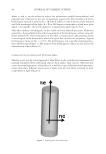
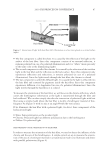
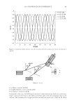
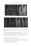
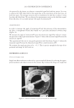
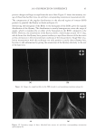
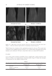
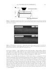
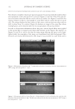
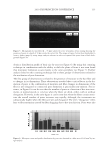
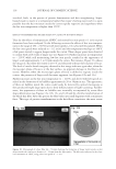
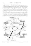
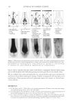
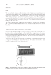
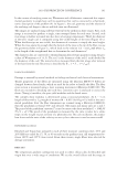
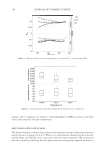
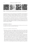
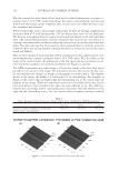
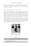
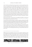
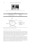
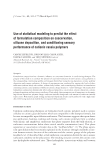
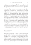
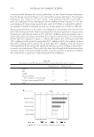
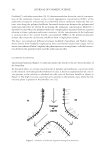
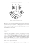
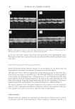
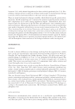
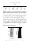
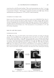
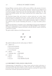
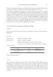
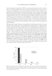
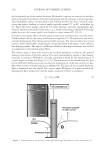
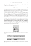
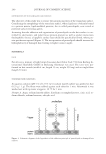
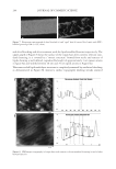
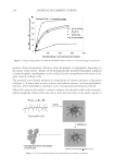
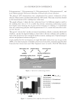
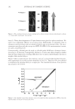
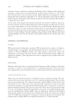
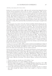
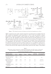
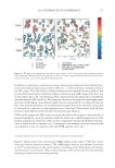
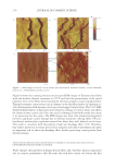
JOURNAL OF COSMETIC SCIENCE 110 applications. Interest on these pores arose from a microscopic investigation made on hair bundles obtained from donors who frequently use hot irons. The investigation revealed that a large number of fi bers from these donors contained a high number of pores. Pores in hair have already been reported by various authors, although, the pores found by these researches does not seem related to hair treatment with hot irons (8–10). In the result section it will be shown that, pores and voids of various sizes, can actually be re-produced in the laboratory when hair is exposed to heat under various controlled conditions. METHODOLOGY The method used to detect the presence of pores in the hair fi bers was by Microscopy using a Hi-Scope from Hirox Model KH-3000. The hair was of Brown European Wavy Virgin Hair from International Hair Importers ~ 8 inches long. Various treatment cycles of hot ironing were applied to hair tresses ~ 0.5 g in weight. The use of this amount of hair allowed the hair fi bers to evenly spread when placed under the hot-iron jaws and ensured that most of them were in contact with the iron surface. Also, whenever needed, cycles of hot ironing were applied to single hair fi bers with a mini-hot iron to confi rm observations. Each treatment cycle of hot ironing consisted of the following steps: 1) 30 s shampooing with a clarifying shampoo, 2) 30 s rinsing with tap water, 3) Blot drying with a towel for 10 s, 4) Blow drying until the hair dried to ~ 80% of its moisture con- tent, 5) Three hot-iron passes over the hair tresses from root to tip at temperatures rang- ing between 180 and 220°C depending on the requirements. Two hot-iron speeds were used during hot ironing, one considered a normal speed ~ 1.0 in/s and the other one con- sidered a low speed ~ 0.2 in/s, and 6) After this process the hair tresses/fi bers were left at rest at room temperature conditions for three hours before applying the next treatment cycle. The method of pore detection consisted in shifting the plane of focus of the microscope from the hair surface into regions inside the cortex according to the diagram showed in Figure 1. This process allowed the visualization of pores and voids inside the cuticle sheath and cortex. The existence of pores was revealed by the contrast of light scatter- ing patterns produced by a mismatch in the indexes of refraction between empty spaces of pores and cortex. Later, counting of the pore number and measurements of their size was made by image analysis using the software Pax-It from Midwest Information Systems. RESULTS DETECTION OF MICROPORES In Figures 2a and 2b are shown micrographs of a hair fi ber with fi ve treatment cycles of hot-iron at 180°C showing typical micropores inside the hair cortex. Figure 2a displays the image of the hair surface taken without shift in plane of focus this picture clearly shows the topology at the cuticle surface. In principle, by looking at this picture it could be inferred that there is no apparent physical change on the hair after its exposure to hot- iron treatment. However, in Figure 2b it can be seen that when the plane of focus of the
Purchased for the exclusive use of nofirst nolast (unknown)
From: SCC Media Library & Resource Center (library.scconline.org)