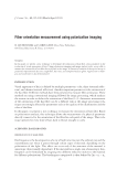
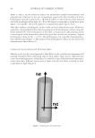
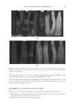
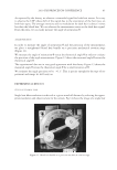
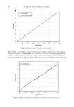
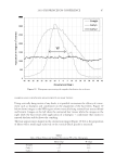
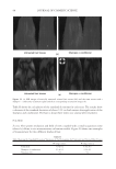
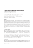
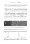
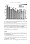
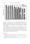
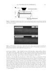
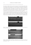
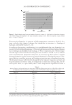
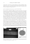
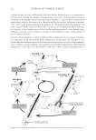
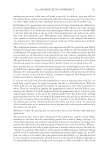
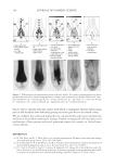
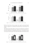
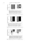
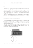
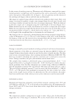
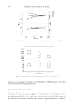
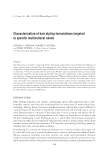
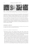
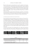
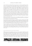
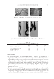
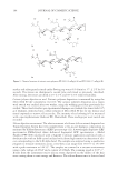
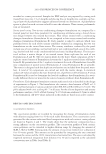
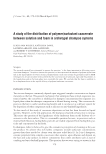
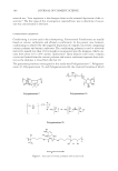
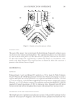
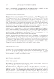
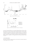
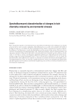
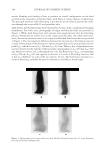
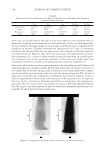
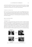
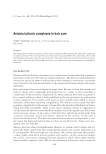
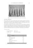
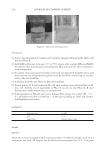
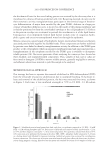
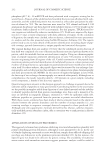
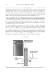
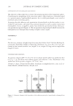
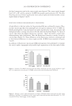
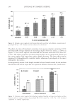
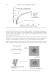
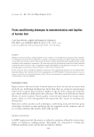
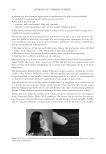
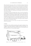
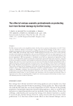
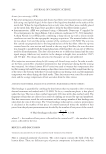
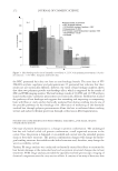
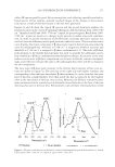
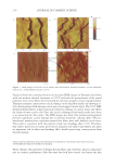
2010 TRI/PRINCETON CONFERENCE 111 microscope is shifted ~ 7 μm towards the center of the hair fi ber, a large number of white dots appears in the hair. These white dots are actually micropores localized inside the hair cortex close to its surface they appear as white dots because the light scattering patterns produced by their empty spaces contrast with the translucency of the cortex. Initially, it was thought that the presence of these pores arose as a consequence of hair chemical treatments such as bleaching or permanent waving. However, a systematic anal- ysis of sets of 200 hair fi bers that were bleached with various degrees of intensity failed to show the appearance of pores. Likewise, sets of hair fi bers from bundles that were treated with permanent waving solutions also did not show the presence of such pores. Further- more, microscopic analysis revealed that only hot-iron treatments were able to create pores in virgin hair fi bers that didn't have any form of damage before treatment. It was diffi cult to measure with precision the size of the micropores however, their apparent sizes as measured by image analysis indicates that their diameter ranges between 0.1 and 1.5 micrometers. Figure 1. Diagrammatic representation of microscope and hair fi ber showing focusing of microscope beam at the hair surface without shift in plane of focus, and at a distance (δ) deep inside into the hair fi ber after the plane of focus was shifted. Figure 2. Micrograph of a hair fi ber (φ ~ 78 μm) taken without shift in plane of focus (2a), and after the plane of focus was shifted towards regions inside the cortex by 7 μm (2b). The hair fi ber was subjected to three hot-iron treatments at 180°C with a hot-iron speed of 1 in/s.

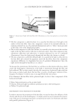
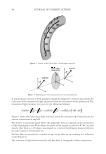
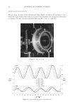
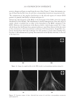
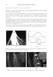
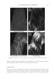
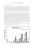
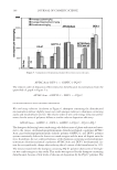
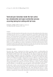
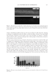
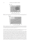
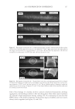
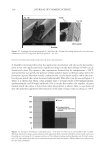
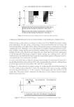
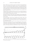
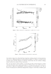
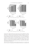
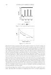
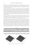
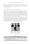
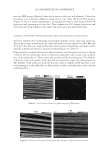
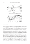
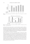
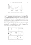
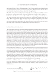
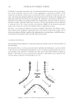
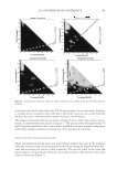
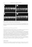
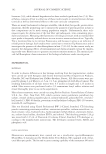
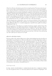
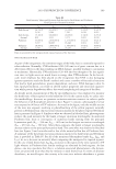
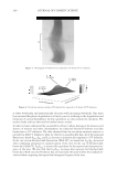
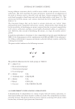
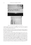
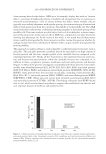
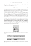
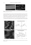
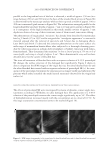
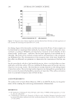
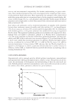
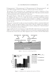
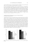
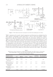
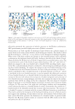
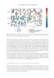
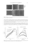
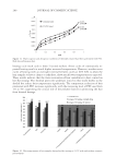
JOURNAL OF COSMETIC SCIENCE 112 EFFECTS OF HOT-IRON TEMPERATURES LOWER THAN 180°C AND NORMAL SPEEDS. Pore density or number of pores per unit area appeared to increase with the number of hot iron treatments at hot iron speeds of 1 in/s (see Figures 3a and 3b). Pores were also ob- served to form in hair that had no cuticle cells on its surface (see Figures 4a and 4b). For instance, Figure 4a shows a micrograph of a hair fi ber with its surface devoid of cuticle cells. The image displays the bare surface of the cortex as this picture was taken with no shift in plane focus. In contrast, Figure 4b displays an image of the same fi ber where multiple pores can be observed after the microscope plane of focus was shifted. In order to obtain more information about the pores, various digital fi lters were applied to the picture fi les to separate pores from other features in the images. For instance, in Figures 5a and 5b it can be seen that by using image fi ltering the pores can be high- lighted while the remainder of the image was transformed into dark background. Once the pores are clearly differentiated in the image it was possible to count them and to Figure 3. Micrographs of hair fi bers (φ ~ 73 μm) subjected to fi ve (3a) and ten (3b) hot-iron treatments at 180°C using a hot-iron speed of ~ 1 in/s. Figure 4. Micrographs (400×) of a hair fi ber (φ ~ 68 μm) devoid of cuticle cells before (4a), and after (4b) the plane of focus was shifted in 5 μm. The fi ber was subjected to fi ve hot-iron treatments at 180°C using a hot-iron speed of ~ 1 in/s.
Purchased for the exclusive use of nofirst nolast (unknown)
From: SCC Media Library & Resource Center (library.scconline.org)