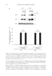
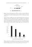
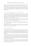
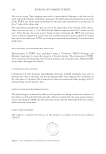
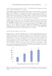
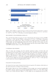
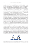
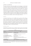
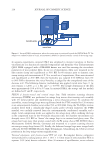
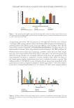
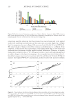
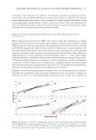
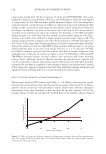
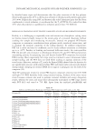
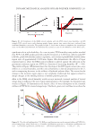
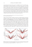
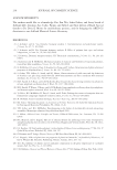
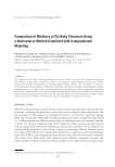
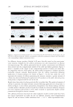
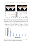
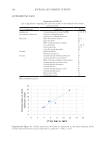
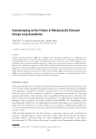
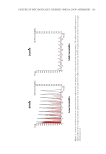
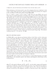
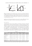
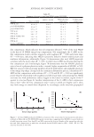
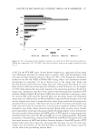
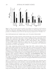
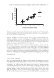
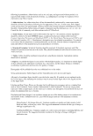
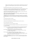
207 Skin Barrier Enhancement by Saccharomyces on the claudin-1 promoting rate up to 108.9 ± 5.5%. SRFF showed the highest promoting effect on claudin-4 production at 12.5 µg/mL. As Stremnitzer reported, Papain-mediated obliteration of claudin-4, ZO-1, and occludin almost doubled TEWL rates 30 minutes after exposure in vivo murine epidermis (22). While water evaporation was significantly reduced, as Malminen observed, there was significantly increased expression of occluding and ZO-1 in the skin after suction blistering within 6 days, and no lipid barrier was present at the same time (23). For TJ-associated marvel proteins, the role of occludin in the barrier function is significant. At SRFF concentrations of 3.13, 12.5, and 50 µg/mL, the protein levels of occludin were analyzed (Figure 4). The protein levels of occludin in epidermal keratinocyte were increased after the SRFF treatment to 107.4 ± 3.4%, 113.5 ± 2.4% (p 0.01), and 115.9 ± 1.9% (p 0.001). The occludin increased in a dose-dependent fashion after the SRFF treatment (Figure 4). At the addition of 50 µg/mL of SRFF content, SRFF showed a significant promoting effect on occludin promoting rate up to 115.9 ± 1.9% (p 0.001). SRFF showed the highest promoting effect on occludin production at 50 µg/mL. SRFF INDUCED THE TJ VERSUS CACL 2 SOLUTION When cultured in medium containing low Ca2+, human epidermal keratinocytes stay undifferentiated and do not develop intercellular contacts (24). The formation of adherens junctions, desmosomes, and then cell stratification can be induced by an increase in extracellular Ca2+ (25). Ca2+ also brings about an increase in occludin, claudin-1, and claudin-4 positive cells. CaCl 2 (3 mmol/L) was used as the positive control group to test the promoting effect on TJs. The results (Figure 5) for claudin-1, claudin-4, and occludin were significantly increased to 111.4 ± 1.1% (p 0.001), 125.5 ± 2.2% (p 0.001), 127.6 ± 4.2% (p 0.001). Our experiment demonstrated that by adding SRFF (12.5 µg/ mL), keratinocyte improvement of claudin-1 and occludin expression (Figure 5) was similar to that induced by a high concentration of Ca2+. Figure 4. Effect of SRFF on the protein levels of occludin in epidermal keratinocytes. Data are presented as the mean ± SE from six experiments. *p 0.05, **p 0.01, ***p 0.001 indicate a statistically significant difference.

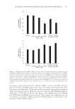
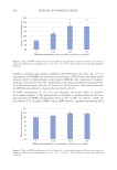
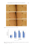
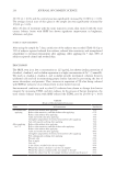
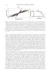
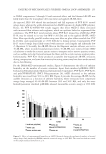
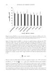
208 JOURNAL OF COSMETIC SCIENCE MEASUREMENTS OF TRANSEPIDERMAL WATER LOSS First, peel-off with adhesive tape for 15 times. Immediately after damage, the skin TEWL is 14.84 (p 0.05), and the TEWL is decreased to 11.12 (p 0.05) after a 10-minute application of the sample lotion. After 10 minutes of sample treatment, the TEWL rate of the sample area was significantly reduced by 25.05%, and that of the control area was significantly reduced by 11.14%. This result means that the yeast essence balance lotion with SRFF instantly helps to repair the skin barrier. COLORIMETRIC MEASUREMENTS After 15 times tape stripping, the skin showed significant redness on both sides and manifested in the increase of the a* value of the skin color. And after the sample treatment, the skin redness on the sample side significantly decreased in 10 minutes, as shown in Figure 6. The skin tone a* value is 8.97 (p 0.05) immediately after damage, and the a* value reduced to 8.32 (p 0.05) after a 10-minute application. In addition, the skin color a* value in the sample area decreased significantly by 7.26% (n = 30, p 0.05) and that in the control area decreased significantly by 3.58%. EVALUATION OF CLINICAL TESTING To evaluate the yeast essence balance lotion’s function against UV radiation, both sides use the same sunscreen in 28 days of clinical testing. The average clinical score of skin brightness in the sample area increased significantly by 16.98%, and the control area increased significantly by 9.92% (p 0.05). The average clinical score of skin whiteness (Figure 7) in the sample area was significantly increased by Figure 5. Effect of SRFF (12.5 µg/mL) and CaCl2 (3 mmol/L) on the protein levels of TJs (claudin-1, claudin-4, and occludin) in epidermal keratinocytes. Data are presented as the mean ± SE from six experiments. *p 0.05, **p 0.01, ***p 0.001 indicate a statistically significant difference.
Purchased for the exclusive use of nofirst nolast (unknown)
From: SCC Media Library & Resource Center (library.scconline.org)