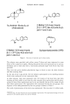
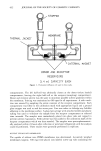
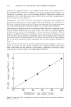
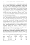
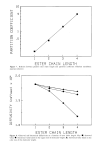
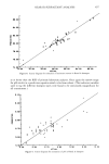
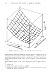
SKIN PLASTICISATION BY 2-HYDROX¾OCTANOIC ACID 399 out all our measurements on whole epidermis with the exception of one experiment where we studied the dependence of human stratum corneum extensibility on environ- mental relative humidity (see below for preparation technique). PREPARATION OF HUMAN STRATUM CORNEUM FOR PENETRATION AND SORPT1ON MEASUREMENT In contrast to the separation procedure described above t•br guinea pig footpads used in mechanical studies, it was necessary to completely isolate stratum corneum layers for penetration and sorption measurements. Substantial sorption of HCA would be expected to any dermis and epidermis present because of their high mass relative to stratum corneum. Thus accurate measurement of sorption to and penetration through stratum corneum would be impossible. Strips of skin obtained with a Davies Simplex electrodermatome from amputated limbs were subjected to the above-described heat separation technique. The resultant epidermal layers were exposed to an established enzyme separation process (11), the remaining layers of stratum corneum being spread onto filter paper and stored in a dessicator. Immediately prior to use, the stratum corneum was rehydrated in water, carefully lifted from the filter paper, and cut to the size required for penetration or sorption. RADIOLABELLED HYDROXYCAPRYLIC ACID 2-Hydroxyoctanoic acid, known as hydroxycaprylic acid (HCA) was synthesised with a •4C atom in the 1-position. Briefly, the synthesis begins with a Grignard reaction involving heptyl bromide and barium carbonate as the radioactive precursor. Dry bro- mine treatment yields the bromo compound which is converted to the hydroxy form by treatment with barium hydroxide. The hydroxy fatty acid is recovered using hydro- chloric acid. After recrystallisation from hexane, its purity measured by thin layer chro- matography was 98%. Solutions of non-labelled HCA were tagged by the addition of small quantities of •4C HCA. EXTENSIBILITY MEASUREMENTS An instrument was designed and made to measure the extensibility of strips of guinea pig footpad epidermis cut to a size of approximately 2 mm in width and 15 to 20 mm in length. These strips were clamped vertically, the upper end to a load cell, the lower end to a motor-driven attachment. The linear rate of stretching was normally 0.01 mm s-1. A chart recorder monitored the output from the load cell against time with a minor correction necessary for creep in the load cell. The whole apparatus together with equilibrating skin samples was enclosed in a sealed chamber in which the temperature and relative humidity were controlled and normally held constant at 20øC and 65%. Six strips of guinea pig footpad epidermis were each immersed in water for 3 hours 20øC, blotted dry, and allowed to equilibrate overnight in the chamber. The following day the extensibility of each piece was measured and expressed as % extension (of the original length at the controlled relative humidity) per 100 g load from the most linear section of the load-deformation graph. The strips were then immersed in an aqueous solution of the test material for 3 hours (o: 20øC, blotted, and re-equilibrated overnight in the chamber. After extensibility measurement the following day, the efficacy of the

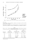
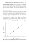
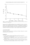
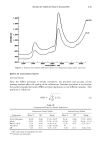
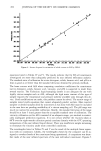
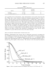
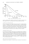
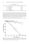
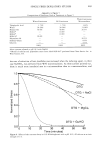
400 JOURNAL OF THE SOCIETY OF COSMETIC CHEMISTS test solution was expressed as the mean ratio (+ two standard errors) of extensibility after test solution treatment to extensibility after treatment with water only. PENETRATION MEASUREMENTS A penetration cell similar to many described in the literature (12) was constructed. The skin layer (2 cm 2 human stratum corneum) was held vertically between glass flanges, thus separating two glass compartments, each with an access port at the top outside edge. Each compartment held about 5 ml solution stirred with magnetic coupling at a rate sufficient to ensure that transport through the solution was not a rate-limiting step. In contact with the outside of the stratum corneum was the donor solution of penerrant, while in contact with the inside was the acceptor solvent, water. The cell was main- tained at a temperature of 30øC, close to that of the skin surface in vivo. Prior to each penetration measurement, the integrity of the stratum corneum was checked by measuring its DC resistance. Isotonic saline was introduced into both chambers. Each side was electrically connected via agar/KC1 bridges to KC1 solution containing calomel electrodes and thence to a high impedance resistance meter. Any membrane whose resistance was below about 40 Kf• was probably faulty and was re- jected. Both compartments were carefully washed out with water before introducing donor and acceptor solutions. The donor solute was radiolabelled and small aliquots were removed at 30-min intervals from the acceptor solution for liquid scintillation counting. Sampling was normally discontinued at 7 hours except in cases of slower penetration rates which were sampled for up to 30 hours. Results were calculated as total amount of solute that had penetrated per 1 cm 2 of skin at a given time from an essentially constant concentration of donor solution (since quantity of solute penetrating total quantity of donor solute). Plotted data showed a gradually increasing amount of penetrating solute tending to a steady-state rate from which the steady-state flux J can be determined. From Fick's first law of diffusion (12), KDAC J - d -- kpAC where K = skin-water partition coefficient D = diffusion coefficient AC = concentration difference across the membrane d = thickness of membrane kp KD - permeability constant kp was calculated for HCA in aqueous solution as a function of solution pH and in the presence of a number of penetration enhancers and structurally related materials. Each study of dependence on one parameter was made, when possible, using skin from one subject to avoid problems of inter-subject variation. All penetration measurements were carried out in duplicate. SORPTION MEASUREMENTS Eight discs of 5 mm diameter were cut from separated human stratum corneum and
Purchased for the exclusive use of nofirst nolast (unknown)
From: SCC Media Library & Resource Center (library.scconline.org)