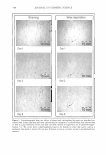
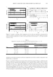
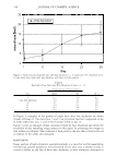
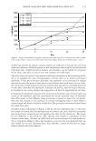
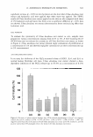
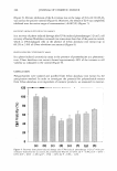
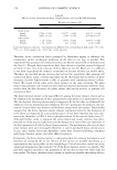
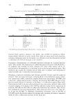
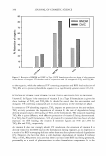
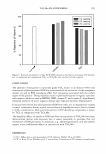
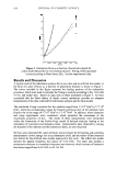
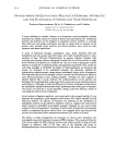
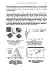
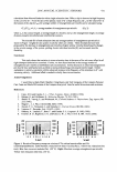
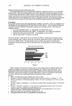
354 JOURNAL OF COSMETIC SCIENCE evaluation can be used (see Figure 5 ). Thus, the determined data is from the same subject, test area, and measurement time as for the assessment of hair reappearance. For a final classification of a depilation method, three parameters, (i) relative regrowth velocity, (ii) depilation closeness, and (iii) irritation level are most important. In this proposed method, all these parameters can be derived from the same set of images in the same study. CONCLUSION Established methods like shaving, plucking, and chemical depilation, and even laser technologies, attain the goal of keeping the skin free of hairs only for a period of time before the hairs reappear. For the quantification of how good a hair removal method is, the determination of at least three parameters is necessary. These parameters are re growth velocity, depilation closeness, and skin irritation. While the time that elapses until hairs reappear on the skin is the most important parameter, the closeness of hair removal as assessed by determining non-removed or incompletely removed hairs is also of great importance to benchmark the quality of hair removal methods. The additional evaluation of the technique's unwanted effects like itch, pain, and erythema, or even injury to the skin, completes the list of the crucial parameters. All three parameters can be assessed in one study close to the daily live situation by using the described study procedure and image-analysis method. REFERENCES (1) E. A. Olsen, Methods of hair removal,]. Am. Acad. Dermatol., 40, 143-155 (1999). (2) J.B. Hamilton, Patterned loss of hair in man types and incidence, Ann. N. Y. Acad. Sci., 53, 708-728 (1951). (3) 0. T. Norwood, Male pattern baldness: Classification and incidence, South. Med.]., 68, 1359-1365 (1975). (4) 0. Braun-Falco and G. P. Heilgemeir, The trichogram. Structural and functional basis, performance, and interpretation, Semin. Dermatol., 4, 40-52 (1985). (5) R. D. Gibbons, V. C. Fiedler-Weiss, D. P. West, and G. Lapin, Quantification of scalp hair-A computer-aided methodology,]. Invest. Dermatol., 86, 78-82 (1986). (6) J. D. Peereboom-Wynia, C.H.Beek, P. G. Mulder, and E. Stolz, The trichogram as a prognostic tool in alopecia areata, Acta Derm. Venereal., 73, 280-282 (1993). (7) M. Saitoh, M. Uzuka, and M. Sakamoto, Human hair cycle, J. Invest. Dermatol., 54, 65-81 (1970). (8) D. J. Van Neste, B. de Brouwer, and W. De Coster, The phototrichogram: Analysis of some technical factors of variation, Skin Pharmacol., 7, 67-72 (1994). (9) R. Hoffmann, TrichoScan: A novel tool for the analysis of hair growth in vivo,]. Invest. Dermatol. Symp. Proc., 8, 109-115 (2003). (10) M. D. Van Neste, Assessment of hair loss: Clinical relevance of hair growth evaluation methods, Clin. Exp. Dermatol., 27, 358-365 (2002). (11) T. Leroy and D. Van Neste, Contrast enhanced phototrichogram pinpoints scalp hair changes in androgen sensitive areas of male adrogenetic alopecia, Skin Res. Technol., 8, 106-111 (2002). (12) D. Van Neste, T. Leroy, and E. Sandraps, Validation and clinical relevance of a novel scalp coverage scoring method, Skin Res. Technol ... 9, 64-72 (2003). (13) M. E. Roersma and G. J. Veldhuis, Proposal and evaluation of a Monte Carlo model for hair regrowth following plucking, Skin Res. Technol., 7, 176-183 (2001). (14) P. Bjerring, H. Egekvist, and T. Blake, Comparison of the efficacy and safety of three different depilatory methods, Skin Res. Technol., 4, 196-199 (1998). (15) R. F. Wagner, Jr., C. A. Flores, and L. F. Argo, A double-blind placebo controlled study of a 5% lidocaine/prilocaine cream (EMLA) for topical anesthesia during thermolysis,j. Dermatol. Surg. Oneal., 20, 148-150 (1994).

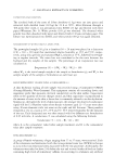
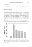
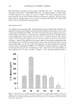
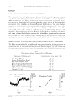
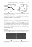
J. Cosmet. Sci., 57, 355-367 (September/October 2006) Cosmeceutical properties of polysaccharides from the root bark of Ulmus davidiana var. japonica SANG YONG EOM, CHAN BOK CHUNG, YOUNG SIL KIM, JONG HEON KIM, KI SOO KIM, YOUNG HEDI KIM, SUN HEE PARK, YONG-IL HWANG, and KI HO KIM, R&D Center, Chamzzone, Taejang 2, Wonju, Kangwon 220-962 (S. Y.E., Y.S.K., J.H.K), R&D Center, Bioland Ltd, Songjeong, Byongchon, Cheonan, Chungnam 330-863 (C.B.C., K.S.K., Y.H.K., S.H.P., K.H.K.), and Division of Food Science and Biotechnology, Kyungnam University, Masan 631-701 (C.B.C., Y.-l.H.), Korea. Accepted for publication May 4, 2006. Presented at the 2005 IFSCC Conference, Florence, Italy, September 19-21, 2005. Synopsis In Korea and China, Ulrnus davidiana var. japonica has been used as a traditional oriental medicine for the treatment of difficulty in urination, skin inflammation, etc. In order to investigate the potential of a polysaccharide extract from Ulrnus davidiana var. japonica as a cosmetic ingredient, we measured its mois turizing effect, photo-induced cytotoxicity, and anti-inflammatory effect. After hydrolysis, HPLC experi ments showed that the composition of the polysaccharide extract was mainly rhamnose, galactose, and glucose. The molecular weight of the obtained V Imus davidiana root extract was 20,000. The intrinsic viscosity was 90 dl/g. In a moisturizing test conducted through the measurement of water loss in a desiccator and of moisture content with a Corneometer CM820, Vlrnus davidiana root extract showed almost the same moisturizing effect as hyaluronic acid. In an assay for inhibition of the H202-activated release of PGE2, IL-6, and IL-8 in normal human fibroblast cell lines, Ulrnus davidiana root extract showed an inhibitory activity of PGE2 release in a dose-dependent manner (up to 85 .9% at a concentration of 0.1 % ). The percent inhibition of the release ofIL-6 was in the range of 45.6% to 64.5% (H202 was used as the positive control). Moreover, the release of IL-8 was completely inhibited in the entire concentration range (0.0025%). In a test of recovery from photo-induced damage after UVA irradiation (3 J/cm2), the cell recovery of human fibroblasts increased to levels two times higher than that of the positive control, which was UVA-damaged cells in the absence of Ulrnus davidiana root extract (up to 60.2% at 3.0% of Vlrnus davidiana root extract). In a photo-induced cytotoxicity assay in the presence of promethazine as a photosensitizer, Ulrnus davidiana root extract showed approximately 48% of the increased cell viability of the control. Therefore, U lrnus davidiana root extract may be useful for the development of a cosmetic ingredient. INTRODUCTION U !mus davidiana var. japonica (Family: Ulmaceae) 1s a deciduous tree that 1s widely Address all correspondence to Ki Ho Kim. 355
Purchased for the exclusive use of nofirst nolast (unknown)
From: SCC Media Library & Resource Center (library.scconline.org)