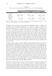
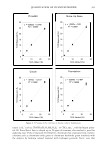
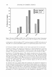
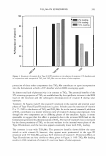
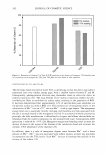
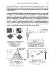
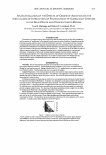
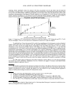
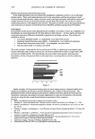
428 JOURNAL OF COSMETIC SCIENCE ROLE OF SODIUM LAURYL SULFATE (SLS) MICELLES IN INDUCING SKIN BARRIER DAMAGE IN THE PRESENCE OF GLYCEROL: DOES MICELLE SIZE MAT TER? Saswata Ghosh and Daniel Blankschtein, Ph.D. Department of Chemical Engineering, MIT, Cambridge, MA 02139 Introduction and Significance Surfactants commonly encountered in skin-care formulations are known to reduce the barrier properties of the skin. It is well-accepted that surfactants have to first penetrate into the skin barrier before they can reduce skin barrier properties. Therefore, if a formulator can minimize surfactant-skin penetration, this should also minimize the ability of the surfactant to reduce the skin barrier properties. Surfactants and other hydrophilic chemicals can penetrate into the skin barrier through aqueous pores in the stratum corneum (SC). [JJ These aqueous pores in the SC provide the primary skin barrier penetration and transport pathway for hydrophilic chemicals which would otherwise not be able to penetrate into the skin barrier through the lipoidal, hydrophobic pathways that exist in the SC. 121 Sodium Lauryl Sulfate (SLS), an anionic surfactant and a model skin irritant, disrupts the skin barrier upon coming in contact with it. SLS monomers self-assemble to form micelles at concentrations above the Critical Micelle Concentration (CMC). We Ill and others l 3 J have observed that the SLS-induced barrier disruption is dose-dependent, and increases with an increase in the total SLS concentration above the CMC. This important observation contradicts the well-accepted Monomer Penetration Model (A,{PM), which attempts to explain surfactant-skin penetration by considering solely the surfactant monomers which can penetrate the skin barrier through the aqueous pores in the SC. The MPM does not account for the possibility that surfactant micelles can also contribute to surfactant-skin penetration, because it considers the micelles to be too large to penetrate through the aqueous pores in the SC. Our recent SLS-skin penetration studies indicate that the aqueous pores in the SC increase in size upon coming in contact with SLS, such that the average pore radius is greater than the SLS micelle radius. Therefore, SLS micelles, contrary to the view put forward by the MPM, are not sterically hindered from penetrating into the skin barrier through these pores. Furthermore, our studies conclusively show that the contribution of the SLS micelles to SLS-skin penetration dominates that of the SLS monomers at concentrations above the CMC, which are typically encountered in skin-care formulations. Therefore, strategies to reduce the contribution of SLS micelles to SLS-skin penetration can si gn ificantly reduce the amount of SLS that can penetrate into the skin barrier and induce skin irritation. In this paper, we have investigated such a practical strategy, using mixtures of Glycerol (a well-known humectant) and SLS, and have found that the addition of Glycerol eliminates almost completely the contribution of the SLS micelles to SLS-skin penetration. Experimental and Theoretical Framework Excised pig skin samples were hydrated in Franz Diffusion Cells (FDCs). Subsequently, the donor compartments of the FDCs were filled with solutions of SLS, SLS+l0 wt% Glycerol, 10wt% Glycerol, and Phosphate Buffered Saline (PBS, the control). The skin barrier properties were quantified by measuring the electrical current across the skin at appropriate voltage signals. In short, the higher the measured skin electrical current for identical voltage signals, the lower is the skin resistance, and hence, the greater is the reduction in the skin barrier properties. The skin permeability characteristics were quantified by measuring the transdermal permeability of mannitol upon exposure to these contacting solutions. Mannitol is a hydrophilic probe that traverses the SC through the same aqueous pores that allow the transport of the current-carrying ions. The amounts of SLS that may penetrate into the skin barrier from the SLS contacting solutions described above were quantified through skin radioactivity assays utilizing 14 C-SLS. For a detailed description of the experimental protocol, see Moore et alY1 A theoretical hindered-transport description ofmannitol and ion transport through the aqueous pores in the SC enabled us to determine: (i) the average aqueous pore radius, and (ii) the aqueous pore number density. For a detailed description of the hindered-transport model, sec Tang et al.l4l Two-Photon Fluorescence Microscopy (TPM) was used to directly visualize the effect of SLS and SLS+ 10wt% Glycerol on excised pig skin samples in FDCs. For detailed descriptions of the TPM apparatus and the skin imaging procedure implemented in our studies, see Yu et al.l5 1 . Dynamic Light Scattering (DLS) was used to determine the average SLS micelle size in SLS solutions with and without 10 wi°/o Glycerol. Surface Tensiometry (ST) was used to determine CMC values for SLS in the presence and in the absence of 10 wt% Glycerol. Results and Discussion The results of our skin radioactivity assays using 14 C-SLS in SLS and in SLS+ l0wto/o Glycerol contacting solutions are shown in Figure 1, which plots the concentration of SLS in the skin barrier (triangles and diamonds) versus the total SLS concentration in the contacting solutions. Clearly, since the triangles, which correspond to SLS concentrations in the skin barrier that are significantly lower than those which correspond to the diamonds, it follows that the presence of Glycerol in the SLS

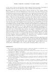
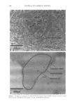
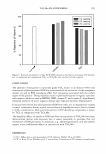
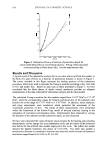
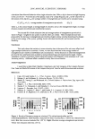
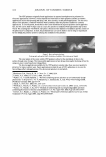
2006 ANNUAL SCIENTIFIC SEMINAR 429 contacting solution significantly reduces the amount of SLS that can penetrate into the skin barrier from the high SLS concentration contacting solutions. The significant difference between the diamonds (or the dashed line) and the triangles (which lie very close to the SLS monomer contribution corresponding to the full line) clearly shows that SLS micelles, which would contribute to skin penetration in the absence of Glycerol, cannot do so in the presence of 10 wt% Glycerol in the contacting solution. Therefore, the addition of 10 wt% Glycerol to the SLS contacting solutions represents a simple, yet useful, strategy to mitigate SLS-induced skin barrier perturbation by preventing the SLS micelles from penetrating into the skin barrier. ? 10 ..--___ ♦_S_L_S ....;. (1_-2_0_ _m_M""") _J.S_L_S ...a (_1-_20_0_m_ M....;.)_+_G""'lyc_e_r_o 1_0_wt'l_Yo_)-----, -� 8 m � 7 II) .c_"' & , CMC o SLS • 8.7 mM ! 5 : .. - -r- - - -- : .. f -- /�-- Micelle Contribution 1 2 L --r --1-- . A § 1 •' • l.... Monomer 8 o.,.�_..__, _________ ---,-------.------,.....I Contribution 50 100 150 200 Total SLS Concentration in the Contacting Solutions (mM) Figure 1. Comparison of SLS-skin penetration induced by contacting solutions of SLS (diamonds) and SLS+ 10 wt% Glycerol (triangles). The error bars represent a standard error based on 6-10 pig skin samples. To understand how Glycerol may prevent SLS micelles from penetrating into the skin barrier, we used our hindered transport model along with our skin electrical current and transderrnal permeability measurements to determine the average aqueous pore radius and the pore number density corresponding to the skin barrier upon exposure to the two solutions considered: (A) SLS and (B) SLS+ 10wt% Glycerol. The average pore radius corresponding to (A) is 33A while that corresponding to (B) is 20A, which is similar to the aqueous pore radius corresponding to the PBS control. In addition, the pore number density corresponding to (A) is twice that corresponding to (B). Using DLS experiments, we measured the SLS micelle hydrodynamic radius corresponding to (A) to be 19.5A and that corresponding to (B) to be 18.0A. Furthermore, our ST studies did not indicate any statistical difference between the CMCs corresponding to (A) and (B). Therefore, these results indicate that Glycerol does not reduce SLS-skin penetration by either: (i) reducing the CMC such that a lower concentration ofSLS monomers can contact and penetrate into the skin barrier, or (ii) increasing the SLS micelle size such that the larger SLS micelles cannot penetrate through the aqueous pores in the SC. In fact, our results seem to indicate that Glycerol reduces the size of the aqueous pores in the SC relative to that of the SLS micelles, which if not reduced, would allow SLS micelles to contribute to SLS-skin penetration. Our TPM studies indicate, through direct skin barrier visualization, that the addition of 10 wt% Glycerol minimizes SLS-induced skin barrier perturbation. Furthermore, the use of Glycerol also appears to minimize SLS-induced keratin denaturation within the comeocytes of the SC. Conclusions SLS micelles can penetrate into the skin barrier and induce skin irritation. The addition of GI ycerol was found to reduce the size of the aqueous pores in the SC, such that the SLS micelles can no longer penetrate into the skin barrier and induce skin irritation in the presence of Glycerol. Therefore, the addition of 10 wt% Glycerol to a SLS contacting solution represents a simple, yet useful, practical strategy to minimize SLS-induced skin irritation. References 1. P.Moore, S.Puvvada, and D.Blankschtein, Journal of Cosmetic Science, 54, 29-46 (2003). 2. G.K.Menon, and P.M.Elias, Skin Pharmacology, 10, 235-246 (1997). 3. L.D.Rhein, F.A.Simion, R.L.Hill, RH.Cagan, J.Mattai, and H.I.Maibach, Dermatologica, 180, 18-23 (1990). 4. H.Tang, S.Mitragotri, D.Blankschtein, and R.Langer, Journal of Pharmaceutical Sciences, 90, 545-568 (2001). 5. B.Yu, K.H.Kim, P.T.So, D.Blankschtein, and R.Langer,Joumal of Investigative Dermatology, 120, 448-455 (2003). Acknowledgements We would like to thank Dr. Sidney Homby and Dr. Yohini Appa from Neutrogena Corporation for useful discussions, and for providing partial financial support for this work
Purchased for the exclusive use of nofirst nolast (unknown)
From: SCC Media Library & Resource Center (library.scconline.org)