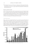
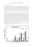
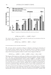
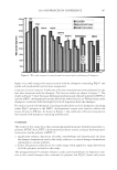
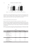
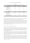
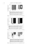
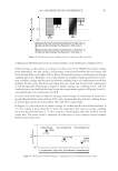
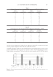
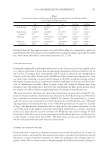
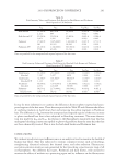
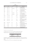
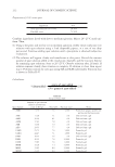
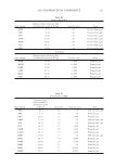
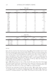
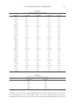
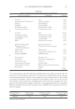
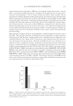
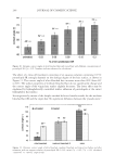
JOURNAL OF COSMETIC SCIENCE 228 WET COMB All products were evaluated on 10-inch virgin brown hair. Two x 2-gram swatches were used for each material tested, all from the same lot. All swatches were wet with 25°C water and one gram of test material was used for each swatch. Swatches were washed and then rinsed for at least one minute per swatch. Wet comb evaluation was then performed. No blow-drying of hair was done. All swatches airdried then the dry comb evaluation was performed once hair was completely dry. Scale used is 1 to 5, 5 being the best. Used for wet and dry combing. Sample/Evaluation Wet comb Rinse-off Clean feel (Scroop) Shine Residual feel Average Control water only 1.0 3.0 2.0 2.0 2.0 2.0 FH183D 4.5 4.5 4.5 3.0 3.0 3.9 DRY COMB Sample/ Evaluation Dry comb Dry feel Clean feel/ look Shine Fullness/ manageable Fly-away Residual feel Static Average Control water only 3.0 3.0 2.0 1.0 1.0 1.0 1.0 2.0 1.750 FH183D 4.4 4.5 4.0 4.0 4.4 4.2 3.5 4.0 4.125 SALT TOLERANCE, PH, VISCOSITY, EASE OF FORMULATION, AND EFFECT ON FORMULATION STABILITY The scale used is 1 to 5, 5 being the best, only for salt tolerance, ease of formulation, and effect on formulation stability. Viscosity was tested by using a Brookfi led, LVT, #4 spindle, 12 rpm. Formula/ Evaluation Salt tolerance pH Viscosity, cps Ease of formulation Effect on formulation stability Average FH183D 2.5 5.70 12,000 4.0 4.5 3.67 REFERENCES (1) R. G. Pearson, J Am Chem Soc., 85, 335 (1963). (2) E. Lucassen and D. Giles, J. Colloid Interface Sci., 81(1), 150–157 (1981). (3) A. J. O’Lenick, J. Surfact. Deterg. 3(2), 229 (2000). (4) G. Kume, M. Gallott, and G. Nunes, J. Surfact. Deterg., 11, 1–1 (2008).

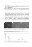
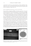
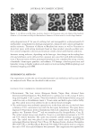
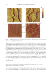
J. Cosmet. Sci., 62, 229–236 (March/April 2011) 229 Proteomic analysis of hair shaft and nail plate ROBERT H. RICE, Department of Environmental Toxicology, University of California, Davis, CA 95616-8588. Synopsis The protein components of living cells in the hair follicle are amenable to study by standard molecular biolog- ical techniques, but identifying those in the hair shaft has been problematic until recently. Most of the protein, primarily keratins and keratin associated proteins, can be extracted under denaturing conditions, but 15-20% is intractable due to transglutaminase-mediated cross-linking. Shotgun proteomics now permits identifying 300 constituents of the isopeptide cross-linked proteome and even certain post-translational modifi cations. The proteins originate from all the intracellular compartments, indicating that the cross-linking process makes effective use of available resources to produce structures with great mechanical stability. Knowing this proteome provides a foundation for correlating defects in hair shaft structure with protein defi ciencies. Such investigations can be extended to mouse models of aberrant pelage hair. Thus, inbred mouse strains can be distinguished by their hair proteomes, raising the possibility of similar variation in the human population. The nail plate is also amenable to this shotgun proteomic approach. Providing discrete and noninvasive sam- pling of the human proteome, these epidermal appendages could have diagnostic utility for certain disease states. BACKGROUND Mature corneocytes in hair shaft, nail plate and epidermal callus are designed by nature to resist external physical stress and chemical exposures. They are comprised largely of kera- tin and keratin-associated proteins surrounded by an envelope of cross-linked protein. While the majority of protein is extractable from these corneocytes under strongly dena- turing conditions, a substantial fraction (15-20% in the case of hair) resists solubilization due to considerable transglutaminase-mediated isopeptide bonding. An inability to sepa- rate constituent proteins has prevented their identifi cation until very recently. Microscopy of the structures has revealed important physical features, and immunohistochemical studies of developing regions bordering the mature ones has permitted identifi cation of major components. The powerful approach of targeted gene ablation provides comple- mentary information on structural and developmental defects. Now that mass spectrome- try, coupled with database searching of peptide masses, permits identifi cation of proteolytic fragments of complex protein mixtures, the identities of the components of the cross- linked material are now being revealed. Studies by pioneering dermatologists a half century ago pointed to a particularly resistant structure at the outer boundary of corneocytes in the callus layer of the skin (1,2). Similar features are seen in the nail plate, where the interlocking borders of adjacent cells provide
Purchased for the exclusive use of nofirst nolast (unknown)
From: SCC Media Library & Resource Center (library.scconline.org)