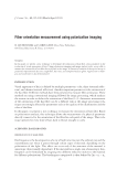
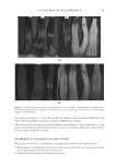
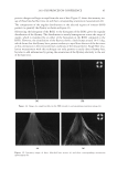
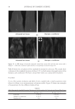
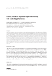
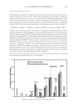
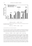
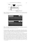
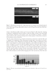
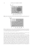
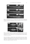
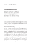
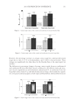
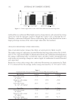
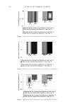
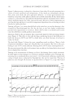
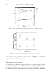
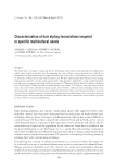
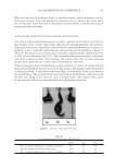
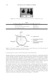
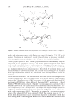
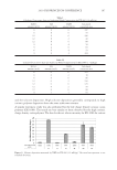
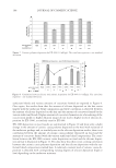
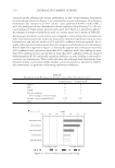
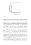
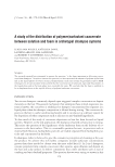
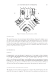
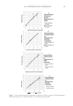
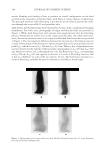
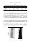
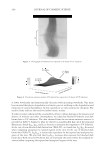
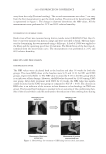
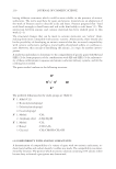
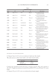
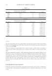
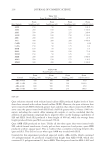
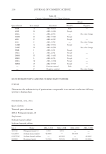
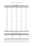
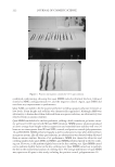
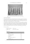
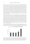
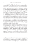
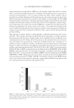
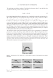
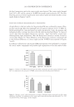
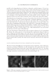
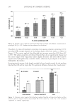
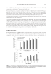
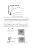
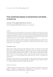
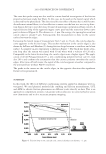
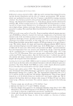
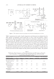
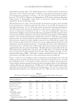
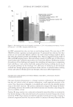
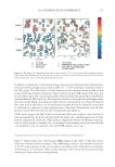
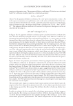
JOURNAL OF COSMETIC SCIENCE 198 where one can visually observe the region of the tress where hot iron treatment was ad- ministered, resulting in the formation of a dark yellow hue. In the case dark brown hair, we do not observe a visually signifi cant color change, probably because it is masked by the absorption of melanin. Thermal treatment was administered for 1 min. of continuous treatment. For this particular hot iron application, such a length of treatment might be considered extensive. However, this time-scale was meant to be representative of cumu- lative treatments (i.e. a series of short treatment protocols) providing an overall equiva- lent treatment time to the continous treatment. In fact, previous studies show that cumulative treatment is actually more damaging than continuous treatment (7). Data extracted from the excitation-emission matrices for thermally treated Piedmont and dark brown hair are provided in Table III. For dark brown hair, we observe a decrease in the residual Trp levels while the IKyn and I509 bands are statistically similar when compar- ing the thermally exposed region of the tress with the unexposed portion. The calculated peak ratios reveal the same information. In Piedmont hair, thermal exposure results in Trp loss, degradation of kynurenines, and an increase in the intensity at I509. As expected, the ratio, ITrp/IKyn, decreases in thermally exposed hair as compared to the unexposed re- gion of the tress. In contrast, there is an increase in I509/IKyn in thermally exposed Piedmont hair. We suspect that the formation of yellow coloration in thermally exposed hair is re- sponsible for the observed increase in this ratio. Table II Peak Intensity Values and Pertinent Peak Ratios for Dark Brown and Piedmont Hair Exposed to Chemical Relaxer Treatment ITrp IKyn I509 ITrp/IKyn I509/IKyn Dark brown- relaxer 19,150 ± 636 108,000 ± 2,828 26,650 ± 1061 0.177 ± 0.001 0.247 ± 0.003 Piedmont- relaxer 26,150 ± 1,485 2,155,000 ± 35,355 728,700 ± 28,284 0.012 ± 0.000 0.672 ± 0.002 Figure 4. Photographs of thermally exposed (A) Piedmont and (B) dark brown hair.

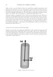
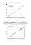
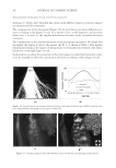
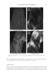
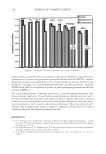
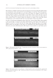
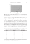
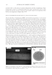
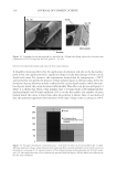
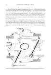
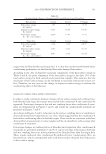
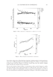
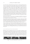
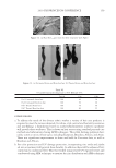
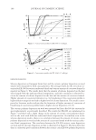
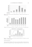
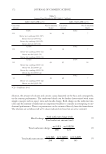
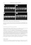
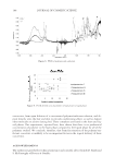
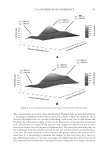
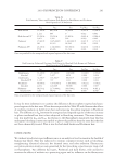
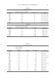
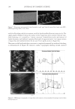
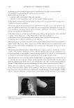
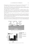
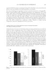
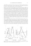
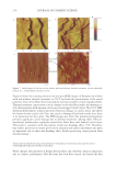
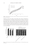
2010 TRI/PRINCETON CONFERENCE 199 PHOTODAMAGE OF HAIR As part of the integument, the outermost organ of the body, hair is constantly exposed to solar radiation. Normally, UVB radiation (290–320 nm) is of great concern due to it deleterious effects in the skin resulting in DNA damage, possibly leading to carcinomas or melanoma. Historically, UVA radiation (320-400 nm) was thought to be less signifi - cant since its light waves are much lower in energy than UVB radiation. In the last de- cade, more emphasis has been placed on the recognition that UVA is also damaging (greater quantities reach the Earth’s surface) and causes a number of free radical reactions that lead to lipid peroxidation, protein degradation, and even, DNA damage in skin. In hair, lipid peroxidation, can result in altered surface properties and structural organiza- tion while protein degradation affects the overal morphological integrity of the fi ber. As already noted, measurement of Trp by spectrofl uorescence was employed to monitor the health state of hair exposed to solar radiation (10). In the current study, we utilize sim- ilar methodology however, we generate excitation-emission matrices in order to probe the behavior of all fl uorophores present in hair. Figure 5 contains a photograph of a hair tress exposed to 96 hours of UV radiation. As noted in the fi gure, only the middle section of the tress was exposed, resulting in photobleaching of the yellow pigment naturally present in Piedmont hair. An excitation-emission matrix for the photo-exposed Piedmont hair is presented in Figure 6. Similar to what we observed in bleached hair, photoexposure reduces the peak intensity for the bands at longer excitation wavelengths. In untreated Piedmont hair, there is convergence of numerous bands starting from the principal kynurenine peak (λex = 394 nm, λem = 465 nm) to the longest excitation wavelength (λex = 450 nm, λem = 509 nm). After photo-exposure, the bands at longer wavelengths decrease relative to the principal kynurenine band. This same effect was observed in bleached hair (see Figure 3) and coincides with a loss of the natural yellow hue of Piedmont hair. A summary of the data from excitation-emission matrices for dark brown and Piedmont hair is provided in Table IV. For all of the monitored fl uorophore bands (ITrp, IKyn, and I509), we see a decrease in intensity when comparing the unexposed to exposed regions. In the case of dark brown hair, ITrp/IKyn increases in the region of the tress exposed to UV light whereas in Piedmont hair similar readings are obtained for both regions. At fi rst glance, one may speculate that this is a melanin-associated phenomenon—the kynure- nines are protected more than Trp by melanin in dark brown hair. In terms of absolute absorbance, we would expect the contrary to be true since melanin absorbance is greater Table III Peak Intensity Values and Pertinent Peak Ratios for Dark Brown and Piedmont Hair Exposed to Thermal Treatment ITrp IKyn I509 ITrp/IKyn I509/IKyn Dark brown 24,550 ± 2,616 103,050 ± 5,586 33,250 ± 7,283 0.239 ± 0.038 0.321 ± 0.053 Dark brown- thermal 15,100 ± 849 108,000 ± 4,243 29,650 ± 4,172 0.140 ± 0.002 0.276 ± 0.049 Piedmont 44,600 ± 5,657 2,030,000 ± 98,995 1,275,000 ± 63,640 0.022 ± 0.001 0.621 ± 0.062 Piedmont- thermal 18,900 ± 1,838 1,800,000 ± 127,279 1,540,000 ± 28,284 0.010 ± 0.000 0.858 ± 0.076 Data are provided for the unexposed and exposed regions of the hair tress.
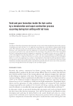
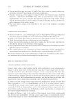
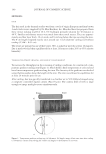
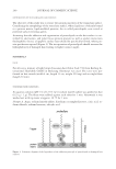
Purchased for the exclusive use of nofirst nolast (unknown)
From: SCC Media Library & Resource Center (library.scconline.org)