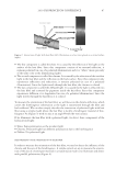
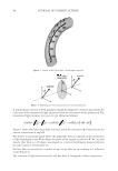
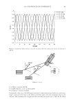
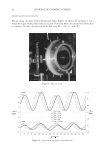
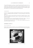
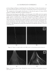
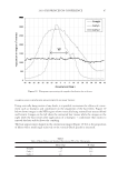
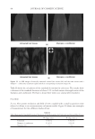
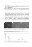
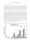
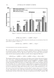
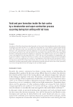
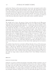
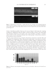
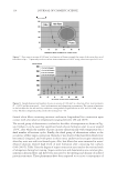
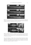
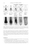
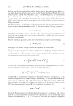
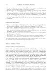
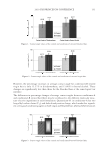
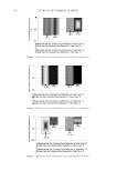
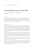
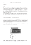
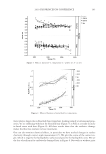
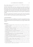
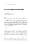
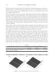
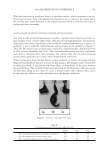
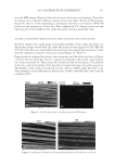
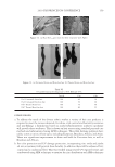
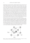
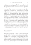
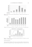
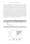
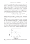
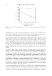
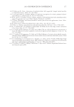
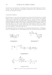
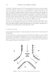
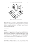
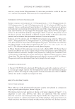
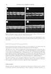
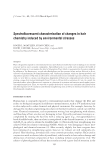
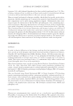
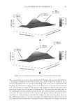
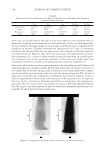
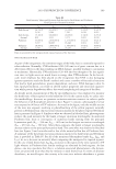
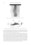
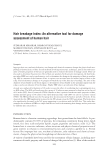
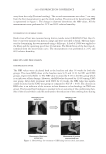
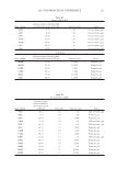
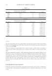
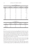
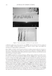
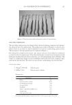
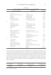
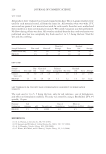
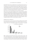
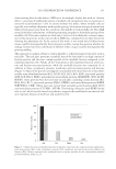
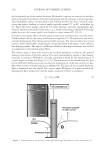
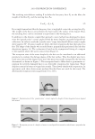
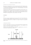
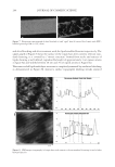
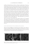
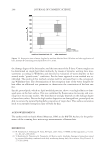
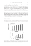
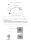
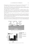
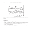
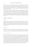
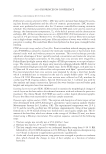
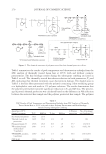
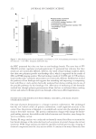
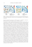
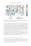
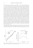
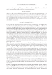
JOURNAL OF COSMETIC SCIENCE 244 and after bleaching and after treatment with the lipid-modifi ed keratins respectively. The upper graph of Figure 8 shows the surface of the virgin hair after cosmetic relevant stan- dard cleansing it is covered by a “mosaic structure” formed from stacks and terraces of lipids showing a well-defi ned, reproducible height of approximately 3 nm (green arrows in fi gure 8a) and widths between 20 nm and 70 nm (pink arrows in Figure 8a). This nano-scaled lipid multilayer structure is completely removed by oxidative bleaching as demonstrated in Figure 8b. Instead a surface topography showing streaks oriented Figure 7. Fluorescence micrograph of ultra bleached (a) and “aged” hair (b) treated (for 5 min) with FITC- labelled proteolipid SR (0.15%, m/m). Figure 8. SFM images (topography) of virgin hair with cosmetic relevant standard cleansing (a) and of ultra bleached hair (b).

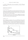
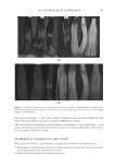
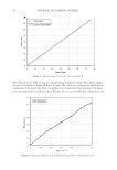
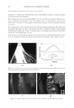
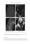
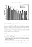
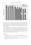
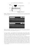
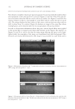
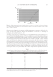
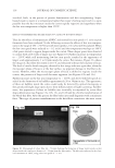
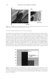
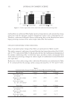
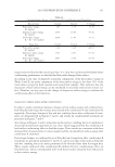
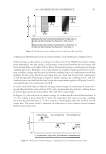
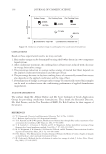
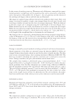
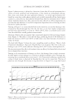
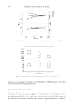
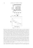
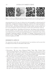
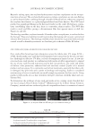
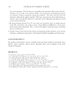
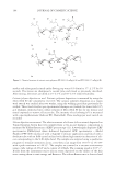
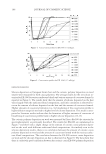
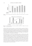
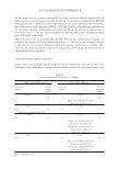
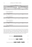
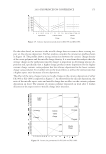
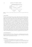
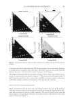
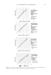
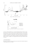
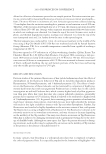
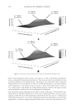
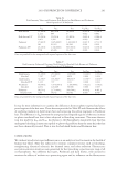
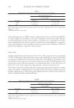
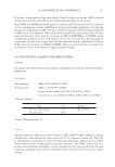
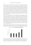
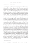
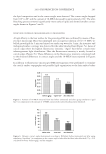
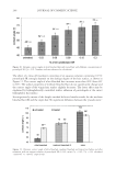
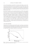
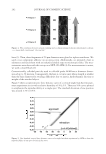
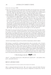
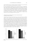
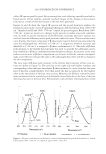
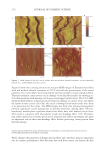
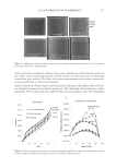
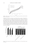
2010 TRI/PRINCETON CONFERENCE 245 parallel to the longitudinal axis of the hair is observed a width of approx. 350 nm (in a range between 200 nm and 500 nm) at the basis of the streaks (black arrows in Figure 8b) is determined by the nanoscope analysis while at their top ends a width of approx. 100 to 200 nm is measured (pink arrows in Figure 8b). The indentations arranged parallel to the longitudinal fi bre axis have depths of approx. 7 nm to 10 nm (green arrows in Figure 8b) as a consequence of the high resolution in this study, smaller cavities of approx. 3 nm depths are observed on top of these striations, some of them round, some more oblong. The phenomenon of longitudinal “striations” has already been described for mammalian hair by J. Smith (17) in 1997 and he assigned this “woodgrain appearance” to exocuticle material exposed after the removal of epicuticle and A-layer due to damaging effects. Later Swift and Smith (7) detected this striation phenomenon on the outer surface of a wide range of mammalian keratin fi bers afore subjected to a thorough cleansing proce- dure by either sonication in sodium dodecyl sulphate or Soxhlet extraction with chloro- form/methanol. They determined the striations “to have a lateral spacing of 350 nm, to be of convex profi le, and rising to a height of approx. 9 nm”. These dimensions fi t very well to those detailed above for the cuticle of bleached hair. The rinse-off treatment of bleached hair with an aqueous solution of 0.15% proteolipid SR changes the surface structure of the damaged hair signifi cantly. Figure 9 shows in direct comparison the SFM images of the virgin hair (a), the ultra bleached hair (b) and the ultra bleached hair treated with an aqueous solution of proteolipid SR (c). In conse- quence of the proteolipid treatment the striations are no longer visible and substituted by patterns which rather resemble the multi layered structures observed for the virgin hair surface (a). EFFECTS OF PROTEOLIPIDS ON THE PHYSICAL PROPERTIES OF THE HAIR SURFACE The effects of proteolipid SR were investigated by means of dynamic contact angle mea- surements according to Wilhelmy on ultra damaged hair. The application of a 0.03% solution of the proteolipid increases the contact angle signifi cantly up to 54°. The effect of hydrophobization increases linearly with the concentration up to a level of 0.15%. At this stage a saturation concentration seems to be reached (Figure 10). Figure 9. SFM image (topography) of a) virgin hair, b) ultra bleached hair, and c) ultra bleached hair treated with an aqueous solution of proteolipid SR (0.15%, m/m).
Purchased for the exclusive use of nofirst nolast (unknown)
From: SCC Media Library & Resource Center (library.scconline.org)