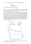
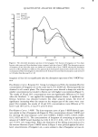
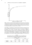
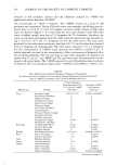
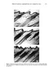
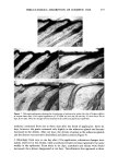
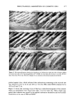
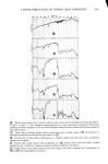
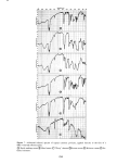
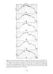
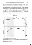
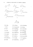
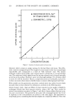
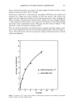
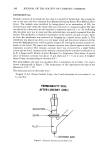
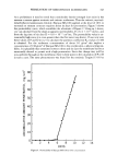
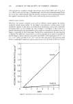
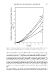
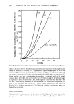
266 JOURNAL OF THE SOCIETY OF COSMETIC CHEMISTS Methods of measuring percutaneous absorption have been reviewed by Ainsworth (13), Barr (14), Tregear (15), Grasso and Lansdown (16) and Idson (17). In this study, the technique of microautoradiography was employed together with that of whole body autoradiography developed by Ullberg (18). Simultaneously, primary irritation potentials were observed histologically on the same substances. In addition, intradermal metabolic fates of the oils were also examined radiochromato- graphically. MATERIALS Isopropyl myristate-•4C (•4C-IPM), decanoxy decane-•C (•C-DD) and 2-hexylde- canoxy octane-•C ( TM C-HDO) were synthesized from 1-•4C-myristic acid, 1-1•C-decyl bromide and 1-•C-octanol respectively in our laboratory. The above starting materials, glyceryl tri-(oleate-1-•C) (•C-GTO) and 1-•4C-octadecane (•C-OD) were obtained from Daiichi Pure Chemicals. These labelled compounds were chemically and radio- chemically pure as checked by thin layer chromatography using several different solvent systems and scanning with a radiochromatogram scanner Aloka TLC-2D. Specific activities of these five oils were adjusted approximately to 0.2/•Ci/mg suitable for this study. An oil-containing hydrophilic ointment was prepared in the following formula: •C- labelled oil 5%, white petrolatum 30%, stearyl alcohol 15%, propylene glycol 12%, sodium lauryl sulfate 2% and distilled water 36%. METHODS WHOLE BODY AUTORADIOGRAPHY WITH HAIRLESS MICE Male hairless mice (hr/hr strain) weighing 25 g (average) were used. Applied to the dorsal skin of animals under occlusion were 0.01 ml of the radioactive oils on 2.5 cm diameter Japanese papers backed with Lumirror © film (Toray Industries, Inc.) or 50 mg of the oil-containing hydrophilic ointments on 2.5 cm diameter filter papers (Toyo Roshi Co., Ltd.). The treated areas of skins were covered with 3M Co.'s Micropore sur- gical tape. The mice were anesthesized with diethyl ether and immersed immediately in a dry ice- acetone bath (-78øC) at different intervals (1, 6, 24 and 48 hr). Subsequently, whole body autoradiography was carried out according to the Ullberg method (18). Forty sagittal sections adhering to Scotch © Tape No. 810 (Sumitomo-3M Co., Ltd.) were prepared with a Jung type microtome from Yamatokoki Co., Ltd., in a freezing room (- l 5øC). The sections were allowed to dry in that room, then brought into contact with Sakura X-ray film Industrial Type N (Konishiroku Photo Ind. Co., Ltd.), and exposed for 40 days. The film development was according to the usual procedure recommended by the manufacturer. MICROAUTORADIOGRAPHY WITH GUINEA PIGS Male guinea pigs (Hartley strain) weighing 340 g (average) were used. The hair on the dorsal region of animals was removed with a hair clipper and an electric shaver one day

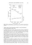
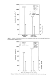
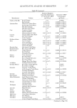
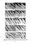
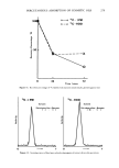
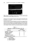
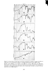
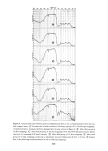
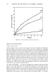
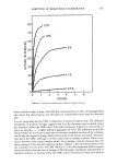
PERCUTANEOUS ABSORPTION OF COSMETIC OILS 267 prior to topical application. The radioactive oils (0.01 ml on 2.0 cm diameter Japanese papers backed with Lumirror © film) were applied to the clipped areas of skins. These treated sites were covered with Micropore © surgical tape. The animals were sacrificed at 6 and 24 hr after application. The skins were excised and f•Ozen immediately by immersion in hexane cooled with dry ice-acetone. Using aJung type microtome, 20/zm frozen sections were cut and dried in a freezing room (- 15øC). After drying the sections were transferred onto the glass slides, and then covered with Fuji autoradiographic stripping films in the dark room. After 50 to 170 days of ex- posure at 5øC, the films were developed according to the usual procedure recom- mended by the manufacturer and the sections were stained in the usual manner with Harris' hematoxylin and mounted. MICROAUTOKADIOGRAPHY WITH ANGORA RABBITS Male Angora rabbits weighing 2.5 kg were used. Two 5 x 6 cm areas were made on the dorsal region of an animal symmetrically with a median line by removing hair with the same manner mentioned above. Then, 0.02 ml of •4C-IPM or •4C-HDO on 3.0 cm diameter Japanese papers were applied to these sites for 2, 6 and 24 hr occlusively. After the animals were sacrificed by air embolism, the treated skins were excised and divided into two pieces parallel to a median line. One was frozen in dry ice for microautoradiography, and the other was fixed in 10% formalin for histological observation. Microautoradiography was undertaken •ccording to the usual method mentioned above. HISTOLOGICAL STUDIES For histological observation, the specimens were embedded in celloidin and paraffin. Serial sections cut at 5/zm mounted on slides were processed through xylol, alcohol to water. The slides were stained in the usual manner with hematoxylin and eosin, and mounted. OBSERVATION OF FATE WITHIN SKIN In the same manner described above, 0.02 ml of•4C-IPM or •4C-HDO were applied to the dorsal skins of Angora rabbits. After 24 hr the •4C-compounds were removed and wiped from the skin surfaces with sanitary cotton. The treated sites were then protected with occlusive dressing during this experiment. At zero, one, three, six and ten days after the removal, the animals were sacrificed by air embolism and the treated skins were excised. Then microautoradiograms and histological specimens were pre- pared by the procedure described above. METABOLIC FATE AFTER INTRADERMAL ADMINISTRATION Angora rabbits, whose hairs had been removed with an electric clipper one day before injection, were injected intradermally in their dorsal skins with 0.05 ml of •4C-IPM (0.14/zCi) or 14C-HDO (0.18/a, Ci). Two rabbits were employed for each substance and six sites were used with each rabbit. At 0, 24 and 72 hr after injection, 1-cm-punch
Purchased for the exclusive use of nofirst nolast (unknown)
From: SCC Media Library & Resource Center (library.scconline.org)