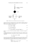
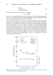
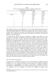
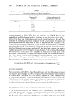
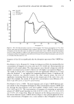
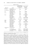
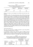
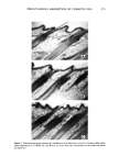
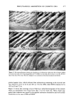
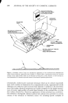
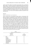
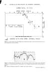
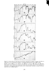
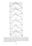
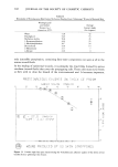
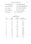
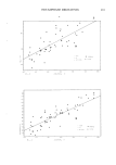
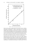
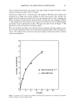
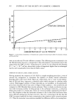
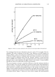
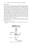
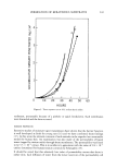
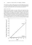
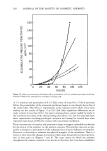
302 JOURNAL OF THE SOCIETY OF COSMETIC CHEMISTS Table II Wettability of Proteinaceous Film Formed by Serous Exudate From "Glistening" Wound of Forearm Skin Wetting Liquid and Surface Average Tension (7•,v) Contact Angle (dynes/cm, 20øC) (0 in degrees) Water 72.8 42 Thiodiglycol 54.0 53 Methylene Iodide 50.8 45 1-Bromonaphth alene 44.6 35 1-Methylnaphthalene 38.7 24 Dicyclohexyl 33.0 14 n- Hexad ecane 27.7 7 n-Decane 23.9 0 only insensible perspiration, containing fatty-ester components not seen at all in the serous wound fluids. In the healing of epidermal wounds, it is primarily the dried films formed by serous exudates (wound fluids) that cover the propagating cells. These cells must move freely as they seek to close the breach of the environmental seal. It becomes important, PROTE]NFtCEOUS E:XLI]OIs::tTE: ON -[t-IIC. I, GE PRISM 0 [0 20 70 SURFtZlCE ' '• ENS'[ON 'D¾NES/CI',1 SLOPE = -0.012 PB]'hI5 h[l'd ,%Yql[ 5'•MBBLS h•}' [NCI dl3E• ]'h STFLq]gdl I•hE FIT '• NOUN]} PRg•OU•EJ] B Y 813 5K]• N 5TR]• PP • NI35 Figure 11. Contact angie data plot characterizing the wettability and adhesive quality of the dried, serous exudate from a "glistening" skin wound.

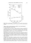
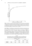
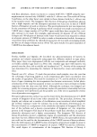
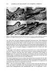
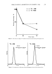
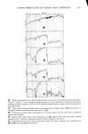
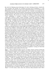
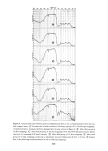
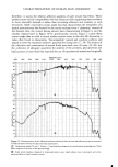
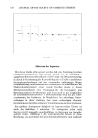
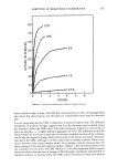
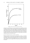
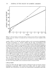
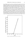
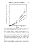
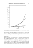
CHARACTERIZATION OF HUMAN SKIN CHEMISTRY 303 therefore, to assess the relative adhesive qualities of such wound fluid films. These qualities must include compatibility with the subjacent cells, supporting their tendency to move smoothly beneath it rather than becoming adherent and resistant to such movement. Table 2 provides contact angle data that characterizes the wettability of a typical proteinaceous film formed by the serous exudate from a "glistening" wound of the forearm skin, the wound having already been characterized in Figure 9, and the exudate characterized in Figure 10 by spectroscopic criteria. Figure 11 plots these contact angle data to yield a critical surface tension value in the mid 20's dynes/cm, a value often found to characterize "biocompatible" natural and synthetic surfaces and argued to have the minimum adhesive potential for living cells (17, 18). The history of the collection and examination of wound fluids goes back over 20 years (19, 20), but the collection of adequate quantities for analysis of the proteins, glycoproteins and lipids present in such fluids has required the use of experimental animals and surgically I FREQUENCY (CM-') 4000 3600 3200 2800 2400 2000 1800 1600 1400 1200 1000 800 650 100 90 80 •7ø 1 •6o z _•$o •4o •3o 2O 10 0 100 90 - • I ................... Figure 12. Demonstration of the different baselines obtained when an internal reflection prism is mounted in different mirror devices for spectroscopic examination. (•) Germanium prism (50 mm x 20 mm x 2 mm) baseline obtained in vertical mirror mounting unit. Note flatter response over most of the spectral range. (•) Same prism baseline obtained in horizontal mirror unit, which allows more convenient and even- pressured skin or liquid cosmetic contact.
Purchased for the exclusive use of nofirst nolast (unknown)
From: SCC Media Library & Resource Center (library.scconline.org)