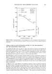
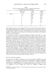
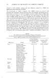
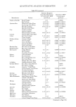
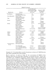
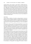
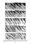
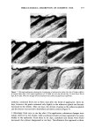
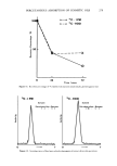
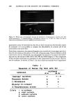
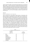
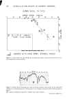
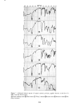
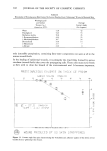
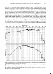
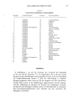
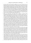
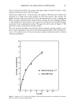
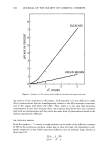
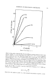
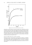
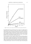
280 JOURNAL OF THE SOCIETY OF COSMETIC CHEMISTS Figure 13. Whole body autoradiograms showing the distribution of radioactivity in hairless mice after topical application of •4C-hexachlorophene in hydrophilic ointment. A) 1 hr, B) 6 hr. After 6 hr, the ra- dioactivity was distributed in the liver, gallbladder and small intestine penetration routes of chemicals into the skin or their interactions with the skin cannot be examined. It is not possible to compare the absorbability of cosmetic oils of low permeability with each other. From these viewpoints, microautoradiography was introduced to investigate and eluci- date the percutaneous absorption of five oils with guinea pigs. The absorbability which could not be seen by whole body autoradiography was found to decrease in the follow- ing order by this method: IPM, GTO, OD, DD and HDO (Figure 2). The comparison of skin irritation potentials was attempted in this experiment by macroscopic observa- tion of erythema. As shown in Table I, the skin irritation potentials were in agreement Table I Response of Guinea Pig Skin with Oil erythema substance 6 hr 24 hr Isopropyl myristate Decanoxy decane n- Octadecane Glyceryl tri-(oleate) 2- Hexyldecanoxy octane Criteria -- no erythema -!- slight erythema -I- moderate erythema -H- severe erythema

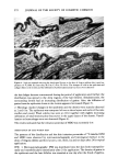
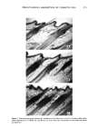
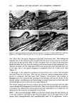
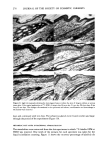
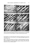
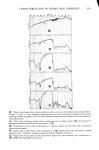
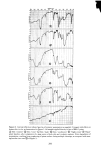
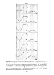
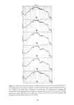
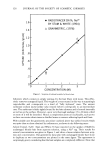
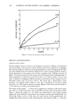
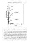
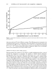
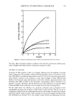
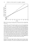
PERCUTANEOUS ABSORPTION OF COSMETIC OILS 281 with the absorbabilities observed by microautoradiography. Therefore it is assumed that the penetration of a material is a necessary condition, though not sufficient in it- self, for the occurrence of irritation. More detailed investigations with Angora rabbits were made on the highest and the lowest absorbed oils to compare them and study the relationship between permeability and irritation. The relationship between accumulation or disappearance of the oils in the skin and occurrence or disappearance of irritation was also an interesting problem. Figures 3 to 6 showed that IPM revealed much more severe irritation than HDO did, though both of these oils penetrated into the skin. As shown in Figures 4 and 6, the ir- ritation patterns of the two were rather different. Namely, IPM induced acanthosis, edematous degeneration of the collagen fibers and changes in the blood vessels, while HDO gave rise mainly to edema in the dermis. Figures 7 and 9 suggested that IPM had a good affinity for the epidermis and the hair follicles and showed a distinct boundary between the dense and sparse distribution of grains, while HDO had a continuous dis- tribution from the epidermis or the sebaceous glands to the adjacent dermis. The grains of both oils increased a little after one day, but gradually disappeared from the skin during the experiment. IPM showed a higher skin irritation potential than HDO did also from the results of macroscopic observation of erythema and microscopic investigation of histology (Figures 8, 10). Since both oils are found to penetrate into the skin, the penetration of a material is not the sufficient condition for the occurrence of irritation. It is evident that the reaction potential of the penetrated material with the skin is the most important factor. The results in Figures 7 to 10 suggested that there existed a time lag from the arrival of the penetrated material at the living cells to the revelation of inflammation. Difference in permeability between the animal species was also observed. The oils were more permeable into the skin of Angora rabbits than into that of guinea pigs. Bartek eta/. showed in their comparison study of animal species (rat, rabbit, pig and man) that drugs penetrated most into rabbit skin and least into human skin, and that the permeability of the skin of miniature swine was the closest to that of human skin (21). IPM was found to penetrate into the skin of miniature swine by microautora- diography in our laboratory (22). According to these findings, this oil is thought to penetrate into human skin also, though IPM is known to generate no erythema on the skin of either miniature swine or man (23). Therefore observation oferythema alone is not sufficient for the safety of cosmetic chemicals. Though the oils that penetrated into the sebaceous glands and the epidermis were seen to eject out of the skin, those in the dermis could not move out in this manner. Did they remain in the dermis or were they carried away by the blood circulation? Micro- autoradiography could not answer this problem clearly. Intradermal administration of the oils partly elucidated the problem showing that IPM became decreased gradually from the dermis HDO tended to remain there, though both were not metabolized in the dermis (Figures 11, 12). From the unpublished data of the authors, IPM was found to be distributed into almost all organs by means of whole body autoradiography when it was injected subcutaneously into mice. The extracts from the liver and kidney were determined as a fatty acid and a triglyceride. On the other hand, HDO was located in the subcutaneously applied site and not distributed into the body organs. These indicated
Purchased for the exclusive use of nofirst nolast (unknown)
From: SCC Media Library & Resource Center (library.scconline.org)