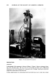
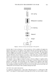
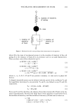
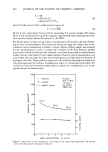
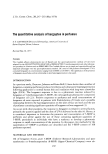
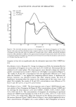
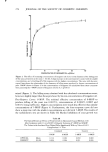
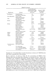
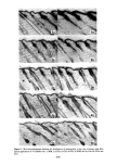
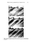
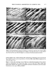
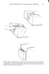
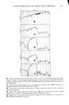
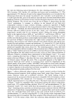
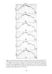
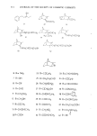
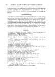
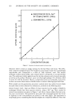
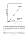
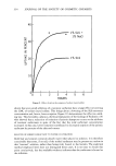
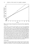
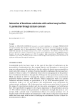
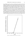
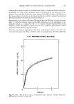
276 JOURNAL OF THE SOCIETY OF COSMETIC CHEMISTS Figure 8. Light micrographs showing the histologic•l feature within the skin of Angora r•bbirs •t v•rious rimes after 26 hr topical application of •C-IPM. A) intact skin, B) zero day, C) one day, D) three days, E) six da•s, F) ten da•s. The chan•es of acanthosis in the e•idermis and edema, vasodilamtion and hemorrhage in the dermis were observed days and continued until ten days. The sebaceous glands were found swollen and large through the period of the experiment (Figure 10). METABOLIC FATE AFTER INTRADEKMAL ADMINISTRATION The metabolites were extracted from the skin specimens in which •4C-labelled IPM or HDO was injected. One tenth of the extracts for each specimen was taken for the liquid scintillation counting. Figure 11 shows the recovery percentage of labelled oils

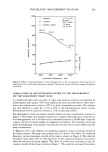
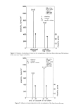
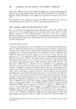
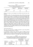
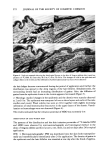
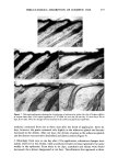
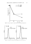
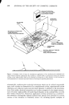
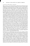
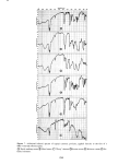
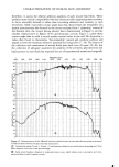
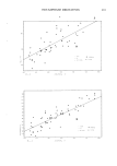
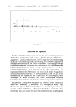
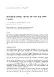
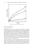
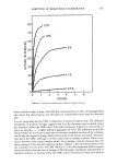
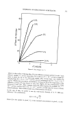
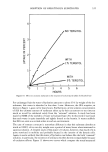
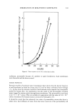
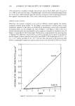
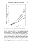
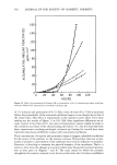
PERCUTANEOUS ABSORPTION OF COSMETIC OILS 277 Figure 9. Microautoradiograms showing the distribution of radioactivity within the skin of Angora rabbits at various times after 24 hr topical application of •4C-HDO. A) intact skin, B) zero day, C) one day, D) three days, E) six days, F) ten days. Note the changes of concentration in the sebaceous glands and dermis plotted against time, which indicated that radioactivity remaining in the injected site was reduced to 43% at 24 hr and 20% at 72 hr for IPM, while HDO remained 42 to 43% at 24 and 72 hr. Figure 12 shows the scanning curves of thin layer radiochromatograms of the extracts where no metabolites were found even after 72 hr for both oils. These results sug- gested that irritation was caused by the applied substances themselves and not by their metabolites in the case of IPM and HDO.
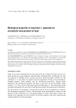
Purchased for the exclusive use of nofirst nolast (unknown)
From: SCC Media Library & Resource Center (library.scconline.org)