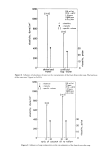
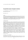
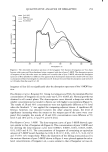
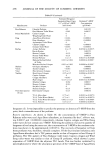
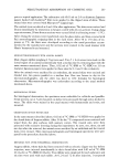
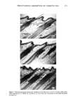
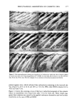
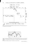
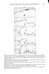
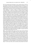
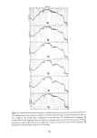
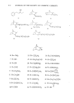
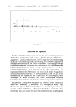
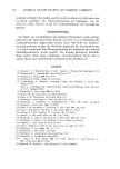
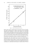
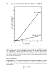
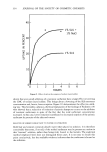
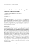
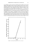
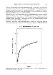
J FIIEQUENCY (CM) ,•O•e 3•00 •ee 2•00 •400 2OOO I•00 I•,00 hmO I •00 mOO •00 •,SO ,•o z Figure 8. Internal reflection infrared spectra of human skin in situ, demonstrating the rapid, noninvasive characterization of both natural and damaged epidermis and the exudates therefrom. (•) "Clean" Baseline for 50 mm x 20 mm x 2 mm germanium prism mounted in horizontal mirror attach- ment, prior to applying subject's forearm. (• Forearm skin of male volunteer, after cleansing with liquid hand soap (see trace A of Figure 5), thorough rinsing, towel drying, and equilibrium with constant condi- tions of "clean room," 72øF, 40% R.H. (• Residue left on prism after skin analysis of (•). (• Forearm skin of male volunteer after 10 "Scotch-tape strippings" of epidermis. (•) Residue on prism following contact analysis of "stripped" skin, in situ. (•) Re-hornified skin of (•, after overnight aging and morning shower.

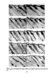
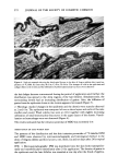
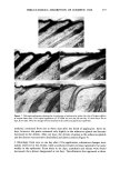
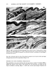
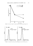
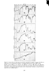
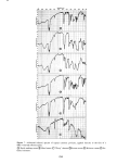
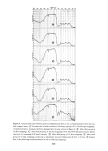
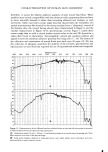
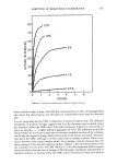
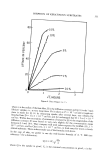
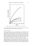
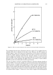
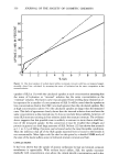
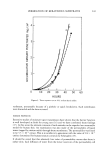
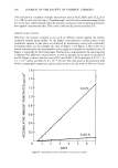
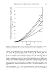
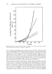
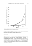
CHARACTERIZATION OF HUMAN SKIN CHEMISTRY 299 subjacent tissues. The final infrared trace of Figure 8 characterizes, once again, the equilibrated skin chemistry of our subject, dried and rehornified for approximately 16 hr following the group of ten Scotch tape strippings. Figure 9 presents spectra collected in another investigation of the changes in skin chemistry and moisture level with depth through the epidermal layers, as those layers were sequentially removed by successive "strippings" with Scotch tape. The uppermost trace of Figure 9 is the internal reflection, infrared spectrum following 20 Scotch tape strippings of the epidermal layers of human skin. This infrared characterization is not significantly different from that shown in Figure 8 after only ten skin strippings. However, as the second trace from the top of Figure 9 shows, following 30 skin-strip- ping operations the epidermal layers under analysis were significantly more hydrated. This is illustrated by the growing infrared absorption band in the region between 3000 and 3600 cm -•. No further significant changes in composition, for example in the lipid to protein ratio, were revealed. The next spectrum shows the consequences of 40 Scotch tape strippings of a forearm skin area, it now becoming clear that the moisture level of these deeper tissues is considerably greater than that nearer the skin surface. The O-H absorption band between 3000 and 3600 cm -• becomes a major band of the spectrum. The fourth trace from the top of Figure 9 characterizes the skin chemistry after 50 Scotch tape applications had stripped away most of the drier epidermal layers, and the fifth trace presents the infrared characterization of the physically wet "glisten- ing" wound produced by 60 such Scotch tape strippings of the epidermis. The final trace of Figure 9 shows the return of the skin chemistry to its equilibrium, normally hydrated state, after the wounded skin zone had healed for six days. Note the reprodu- cibility of both the positions and relative intensities of the infrared absorption bands characterizing the "natural" state of a given subject's skin, as is represented in the three independently obtained traces for that skin included in Figures 8 and 9. EXUDATES AND HEALING OF EPIDERMAL WOUNDS Figure 10 collects internal reflection infrared spectra of the serous exudates collecting at the surface of human skin, in situ, after its wounding to successively deeper layers. The top trace characterizes the exudates of skin "stripped" 20 times with Scotch tape. It is comparable to the spectrum for insensible perspiration and moderate cellular de- bris found for natural or mildly wounded skin as already characterized in Figure 9. The second trace from the top of Figure 10 characterizes the exudates produced after 30 Scotch tape strippings of the epidermal layers. The next trace (third from the top of Figure 10) characterizes the matter extruded from human skin that had been wounded by 40 applications to and sudden removals of Scotch tape from the same skin area. Note, in this 40-layer-deep wound, the beginning of the collection of a wound fluid. The composition of the dissolved components of this fluid, as characterized by infrared absorption bands in the Amide I and Amide II area, is seen to be dominated by proteinaceous substances. The proteins were probably provided by hydrostatic pressure-induced leakage of plasma components through the damaged tissue bed. The infrared traces for human skin wounded to depths of 50 and 60 layers by Scotch tape stripping show increasing amounts of wound fluid collected on the face of germanium prisms during identical contact times, attesting to the increasing compromise of permeability barriers as the skin layers were stripped away. The final trace of Figure 10 shows that, with only a few days of healing, this "glistening" epidermal wound exuded
Purchased for the exclusive use of nofirst nolast (unknown)
From: SCC Media Library & Resource Center (library.scconline.org)