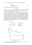
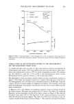
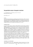
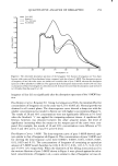
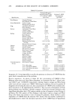
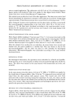
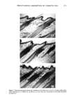
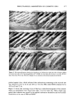
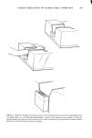
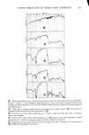
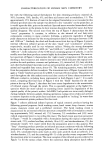
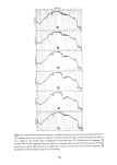
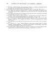
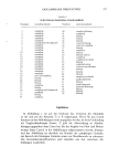
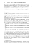
274 JOURNAL OF THE SOCIETY OF COSMETIC CHEMISTS Figure 6. Light micrographs showing the histological feature in the skin of Angora rabbits after topical ap- plication of 14C-HDO. A) intact skin, B) 2 hr, C) 6 hr, D) 24 hr. Acanthosis and edema were observed tion. After that, the grains disappeared gradually from these sites. This finding was assumed due to the removal of the isotope from the skin by the turnover of the epi- dermis and the hair growth. After six days, the grains were not found in the sebaceous glands and after ten days, only the stratum corneum and a part of hair lumina had a dis- tribution of grains. On the other hand, IPM in the dermis decreased abruptly from one to ten days, and so the grains disappeared completely from the dermis at ten days (Figure 7). 2. Histology: in the epidermis acanthosis increased from zero to three days and then decreased from six to ten days. After ten days, however, marked changes were still ob- served as compared with the intact skin. Namely, vacuolization and swelling of Malpighi layer cells remained and swelling of the follicular cells was also found. In the dermis, edema, vasodilatation and hemorrhage increased from zero to three days and remained high and severe from six to ten days (Figure 8). The results showed that irritation reaction proceeded characteristically with the time course though IPM disappeared from the skin. Thus it was assumed that the revelation of irritation was influenced by the sensitivity and the repairability of a living body, not absolutely by the existence or remainder of an irritant. HDO. 1. Microautoradiography: HDO was absorbed deeply along the hair follicles up to the hair matrixes, especially in the sebaceous glands, and in the upper layers of the dermis both transepidermally and transfollicularly after 24 hr application. Such a

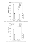
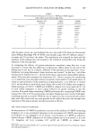
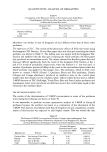
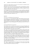
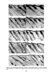
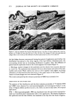
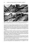
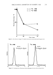
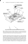
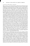
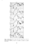
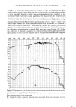
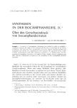
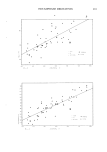
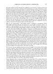
PERCUTANEOUS ABSORPTION OF COSMETIC OILS 275 / •.t -. ,- • ' ., .o}.:..?. .":' ' - -•...:• ..... .., .•.. .•- - :. .•., .,.-....• •..• ..• .... • • .. -t •' .... 4? ,(-•. . '• c•'•'•..?- . 7' •, .... :.:4 './•,• " - ' '...• • • . •" •, ._• ' -'•", •.• •r• - •"•' • •-- . ,.. ,•:•'.:.• • ' •.' • • -• • '•.. ß • . . - ,. -.. - .-, •:• ' . •. .- :.•..., •'. :...•. - • ,. •- "•.: .•-• -- -: . - "' ' •'. ....... ' ' ' '• "• • •-.•"-Qq--s,?•C' •e •" S T':• -• ..--• • • . -.. . '., ß . . •. . - .: • . •'. • .•. -.* ..•-•- •0 ' • . : .. •-•'•,.t • -',• - •: •' , . • .• . •, •' - • , .. •: . ?,%' • . . *-. •.,•,.•.-- -, = . . •. •.•/• '. •- ' ,"• .•.•:' • ... 511 .' •,•.,'•. •,.•. •' *- 'r.•',. *',-,. • . :., • - • ß . *-. *X • . V ' *.-'.•',... ? : * " .... * ' ',) :, , *•**-" .... : V' :X .... '" . Figure 7. Microautoradiograms showing the distribution of radioactivity within the skin of Angora rabbits at various times after 24 hr topical application of •4C-IPM. A) zero day, B) one day, C) $ree days, D) six days, E) ten days. Note the changes of concentration in the sebaceous glands and epidermis tendency continued from one to three days after the finish of application. After six days, however, the grains remained only slightly in the sebaceous glands and became decreased in the dermis. After ten days, the density of grains in the sebaceous glands and the dermis was extremely diminished and almost unseen (Figure 9). 2. Histology: from zero to one day after 24 hr application, edematous changes were mainly observed in the dermis, while acanthosis became increased gradually but quite weakly in the epidermis. From three to six days, acanthosis and edema were found decreased they almost disappeared at ten days. Vasodilatation first appeared at three
Purchased for the exclusive use of nofirst nolast (unknown)
From: SCC Media Library & Resource Center (library.scconline.org)