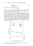
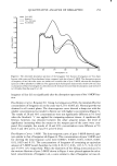
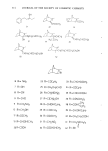
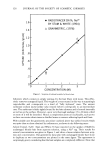
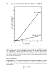
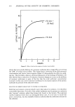
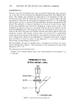
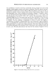
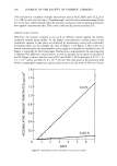
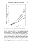
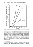
PERCUTANEOUS ABSORPTION OF COSMETIC OILS 267 prior to topical application. The radioactive oils (0.01 ml on 2.0 cm diameter Japanese papers backed with Lumirror © film) were applied to the clipped areas of skins. These treated sites were covered with Micropore © surgical tape. The animals were sacrificed at 6 and 24 hr after application. The skins were excised and f•Ozen immediately by immersion in hexane cooled with dry ice-acetone. Using aJung type microtome, 20/zm frozen sections were cut and dried in a freezing room (- 15øC). After drying the sections were transferred onto the glass slides, and then covered with Fuji autoradiographic stripping films in the dark room. After 50 to 170 days of ex- posure at 5øC, the films were developed according to the usual procedure recom- mended by the manufacturer and the sections were stained in the usual manner with Harris' hematoxylin and mounted. MICROAUTOKADIOGRAPHY WITH ANGORA RABBITS Male Angora rabbits weighing 2.5 kg were used. Two 5 x 6 cm areas were made on the dorsal region of an animal symmetrically with a median line by removing hair with the same manner mentioned above. Then, 0.02 ml of •4C-IPM or •4C-HDO on 3.0 cm diameter Japanese papers were applied to these sites for 2, 6 and 24 hr occlusively. After the animals were sacrificed by air embolism, the treated skins were excised and divided into two pieces parallel to a median line. One was frozen in dry ice for microautoradiography, and the other was fixed in 10% formalin for histological observation. Microautoradiography was undertaken •ccording to the usual method mentioned above. HISTOLOGICAL STUDIES For histological observation, the specimens were embedded in celloidin and paraffin. Serial sections cut at 5/zm mounted on slides were processed through xylol, alcohol to water. The slides were stained in the usual manner with hematoxylin and eosin, and mounted. OBSERVATION OF FATE WITHIN SKIN In the same manner described above, 0.02 ml of•4C-IPM or •4C-HDO were applied to the dorsal skins of Angora rabbits. After 24 hr the •4C-compounds were removed and wiped from the skin surfaces with sanitary cotton. The treated sites were then protected with occlusive dressing during this experiment. At zero, one, three, six and ten days after the removal, the animals were sacrificed by air embolism and the treated skins were excised. Then microautoradiograms and histological specimens were pre- pared by the procedure described above. METABOLIC FATE AFTER INTRADERMAL ADMINISTRATION Angora rabbits, whose hairs had been removed with an electric clipper one day before injection, were injected intradermally in their dorsal skins with 0.05 ml of •4C-IPM (0.14/zCi) or 14C-HDO (0.18/a, Ci). Two rabbits were employed for each substance and six sites were used with each rabbit. At 0, 24 and 72 hr after injection, 1-cm-punch

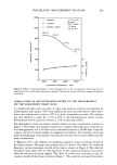
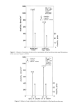
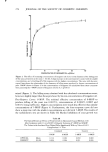
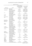
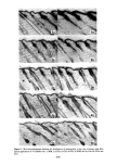
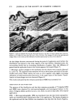
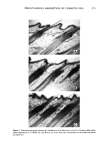
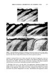
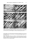
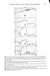
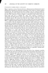
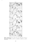
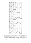
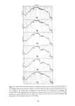
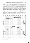
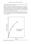
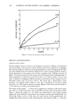
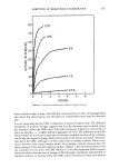
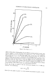
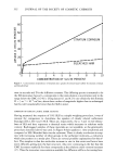
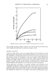
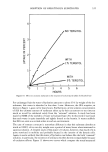
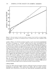
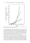
268 JOURNAL OF THE SOCIETY OF COSMETIC CHEMISTS specimens were taken from the injected sites of each animal. Two additional control specimens were obtained from each animal. Metabolites were extracted from each specimen with 4 ml of chloroform in a vial for two days at -20øC in order to avoid decompositions of labelled compounds. After the extract was filtered with cotton wool, the flitrate was dried up quickly. Then the residue was redissolved in 1 ml of chloroform. A 0.1 ml aliquot part of this solution was used for liquid scintillation counting with Aloka LSC-601, and the remainder was spotted on a silica gel plate. After developing, the plate was scanned with a radiochromatogram scanner Aloka TLC-2D in order to identify metabolites. After extraction, the specimen was digested and counted for ra- dioactivity by the method of Petroffet a/. (19). RESULTS WHOLE BODY AUTORADIOGRAPHY WITH HAIRLESS MICE The distribution of •4C-labelled IPM, DD, HDO, GTO and OD was assessed by whole body autoradiography with hairless mice sacrificed at 1, 6, 24 and 48 hr after topical ap- plication. Neither neat oils nor those in hydrophilic ointments penetrated into body organs. The oils were still localized on the applied regions 48 hr after application. The autoradiograms at 48 hr after application of oil-containing ointments are shown in Figure 1. MICROAUTORADIOGRAPHY WITH GUINEA PIGS The distribution of five •4C-labelled oils was observed by microautoradiography in the skins of guinea pigs sacrificed at 6 and 24 hr after topical application. Each oil had a characteristic pattern of distribution in the skin depending on application time. The microautoradiograms of these oils are shown in Figure 2. IPA/I. After 6 hr, the transfollicular penetration was observed, which resulted in the concentration of silver grains into the sebaceous glands. The silver grains are derived from and show the distribution of radioactive substance. The grains were distributed in the stratum spinosum. After 24 hr, the grains were distributed densely in the hair in- fundibula, the follicles, the stratum spinosum and particularly the sebaceous glands. Also, the dermis adjacent to them had a slight distribution of grains. GTO. After 6 hr, the silver grains were distributed from the stratum corneum to the sebaceous glands, but not so marked as those in IPM. After 24 hr, however, the grains spread up to the hair bulges and concentrated considerably in the sebaceous glands. The grains were observed slightly in the dermis under the basal layer and around the hair follicles and the sebaceous glands. OD. After 6 hr, the silver grains were distributed a little from the hair infundibula to the sebaceous glands. After 24 hr, those were concentrated in the sebaceous glands and spread to the dermis around them. DD. After 6 hr, the silver grains did not appear in the skin. After 24 hr, the grains were observed slightly from the hair infundibula to the sebaceous glands. This substance was found to be absorbed at a slow rate.
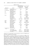
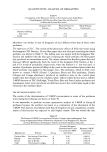
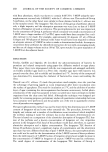
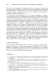
Purchased for the exclusive use of nofirst nolast (unknown)
From: SCC Media Library & Resource Center (library.scconline.org)