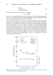
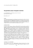
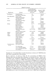
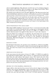
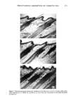
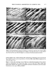
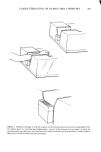
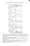
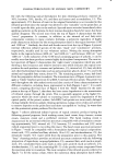
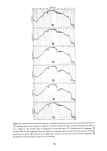
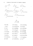
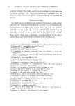
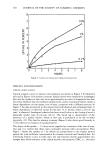
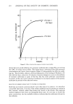
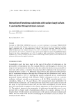
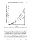
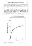
PERCUTANEOUS ABSORPTION OF COSMETIC OILS 271 Figure 3. Microautoradiograms showing the distribution of radioactivity in the skin of Angora rabbits after topical application of 14C-IPM. A) intact skin, B) 2 hr, C) 6 hr, D) 24 hr. Note the concentration in the se- baceous glands and epidermis MICROAUTORADIOGRAPHY AND HISTOLOGY WITH ANGORA RABBITS The distributions and skin irritation potentials of •4C-labelled IPM and HDO were examined by microautoradiography and histological method in the skins of Angora rabbits sacrificed at 2, 6 and 24 hr after topical application. IPM. 1. Microautoradiography: after 2 hr, the silver grains were already observed sparsely in the hair follicles and th• t•pidermis, especially centered in the sebaceous glands. Then the grains became con.•entrated during the period of applicat•.on. The dermis adjacent to the sebaceous glands and beneath the epidermis had an increasing distribution of grains with the time course (Figure 3). 2. Histology: histological changes due to IPM were not intense at 2 and 6 hr. The infiltration of polymorphonuclear leucocytes was observed in the hair follicles. and the collagen fibers under the epidermis were seen to become fine. Also, the infiltration of small mononuclear and polymorphonuclear leucocytes into the upper layers of dermis was slightly observed. After 24 hr, the epidermis was thickened to three or four cell layers. The epidermal cells became swollen and round. Vacuolation around nuclei was also observed. The keratohyalin granules disappeared in a wide area so that the stratum comeurn was thinned. The sebaceous glands became larger. In the dermis, the collagen fibers became fine and marked vasodilatation was seen together with partial hemorrhage (Figure 4). HDO. 1. Microautoradiography: after 2 hr, the silver grains were observed in the se- baceous glands and the adjacent dermis. Then the grains over the sebaceous glands and

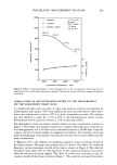
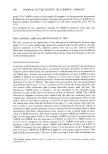
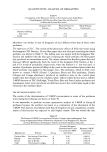
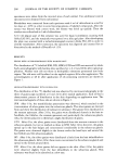
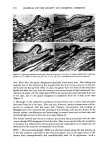
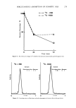
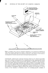
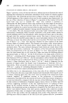
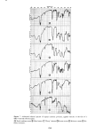
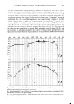
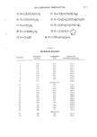
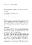
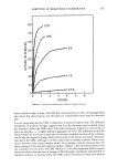
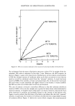
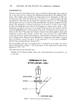
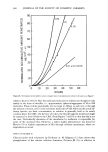
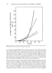
272 JOURNAL OF THE SOCIETY OF COSMETIC CHEMISTS Figure 4. Light micrographs showing the histological feature in the skin of Angora rabbits after topical ap- plication of •4C-IPM. A) intact skin, B) 2 hr, C) 6 hr, D) 24 hr. The changes of cells in the epidermis and collagen fibers in the dermis and the infiltration ofpolymorphonuclear leucocytes were observed the hair bulges became concentrated during the period of application and, further, the distribution was spread to the deep regions of the hair follicles. Simultaneously, the surrounding dermis had an increasing distribution of grains. Also, the diffusion of grains from the epidermis down to the dermis appeared increased (Figure 5). 2. Histology: marked changes in the epidermis and the dermis were scarcely observed at 2 and 6 hr. The epidermis was composed of two or three layers and each cell became swollen and round. Weak edema was seen at 24 hr together with slightly increasing infiltration of small mononuclear leucocytes in the upper layers of the dermis. Vasodi- latation or hemorrhage were not observed (Figure 6). The results indicated that the irritation potential of HDO was extremely low. OBSERVATION OF FATE WITHIN SKIN The patterns of the distribution and the skin irritation potentials of •4C-labelled IPM and HDO were observed by microautoradiography and histological method in the skins of Angora rabbits sacrificed at zero, one, three, six and ten days after 24 hr topical application. IPM. 1. Microautoradiography: IPM was distributed into the skin both transeptider- mally and transfollicularly immediately after 24 hr application. The density of grains in the epidermis and the hair follicles was maximal at one day after the finish of applica-
Purchased for the exclusive use of nofirst nolast (unknown)
From: SCC Media Library & Resource Center (library.scconline.org)