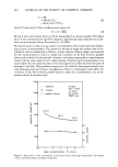
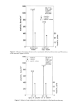
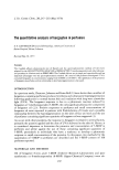
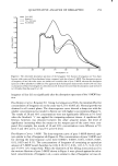
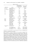
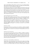
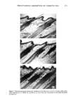
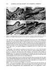
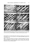
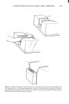
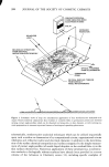
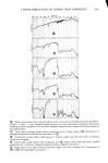
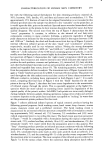
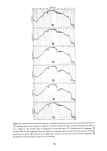
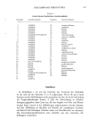
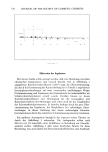
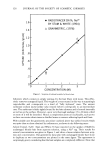
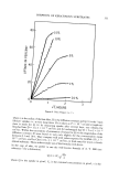
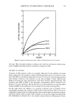
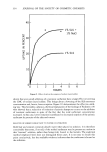
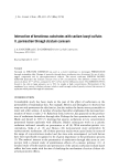
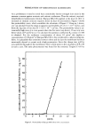
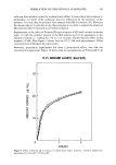
z Figure 10. Internal reflection infrared spectra ofexudates from skin wounded to successively deeper layers. (•) Exudate upon contact with skin "stripped" 20 times with Scotch tape. Compare with skin spectrum, in situ, in Figure 9. (• Exudate after 30 strippings of epidermal layers. ¸ Exudate after 40 strippings. Exudate after 50 skin strippings. Note the absence of lipid absorption bands from this spectrum of purely proteinaceous matter. (•) Exudate from "glistening" forearm wound produced by 60 skin strippings. (•) Exudate from wounded skin area after 6 days of healing. 301

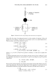
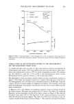
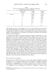
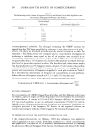
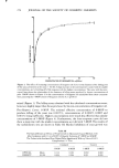
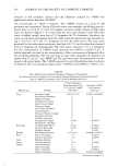
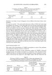
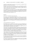
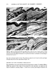
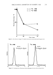
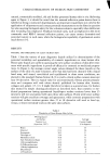
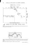
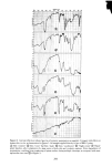
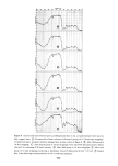
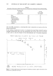
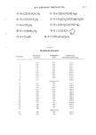
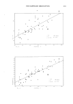
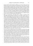
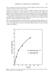
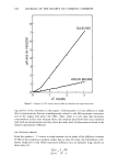
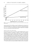
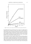
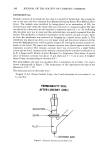
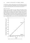
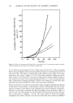
302 JOURNAL OF THE SOCIETY OF COSMETIC CHEMISTS Table II Wettability of Proteinaceous Film Formed by Serous Exudate From "Glistening" Wound of Forearm Skin Wetting Liquid and Surface Average Tension (7•,v) Contact Angle (dynes/cm, 20øC) (0 in degrees) Water 72.8 42 Thiodiglycol 54.0 53 Methylene Iodide 50.8 45 1-Bromonaphth alene 44.6 35 1-Methylnaphthalene 38.7 24 Dicyclohexyl 33.0 14 n- Hexad ecane 27.7 7 n-Decane 23.9 0 only insensible perspiration, containing fatty-ester components not seen at all in the serous wound fluids. In the healing of epidermal wounds, it is primarily the dried films formed by serous exudates (wound fluids) that cover the propagating cells. These cells must move freely as they seek to close the breach of the environmental seal. It becomes important, PROTE]NFtCEOUS E:XLI]OIs::tTE: ON -[t-IIC. I, GE PRISM 0 [0 20 70 SURFtZlCE ' '• ENS'[ON 'D¾NES/CI',1 SLOPE = -0.012 PB]'hI5 h[l'd ,%Yql[ 5'•MBBLS h•}' [NCI dl3E• ]'h STFLq]gdl I•hE FIT '• NOUN]} PRg•OU•EJ] B Y 813 5K]• N 5TR]• PP • NI35 Figure 11. Contact angie data plot characterizing the wettability and adhesive quality of the dried, serous exudate from a "glistening" skin wound.
Purchased for the exclusive use of nofirst nolast (unknown)
From: SCC Media Library & Resource Center (library.scconline.org)