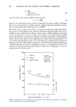
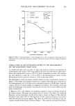
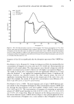
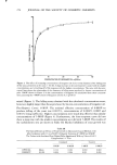
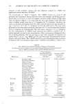
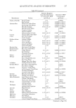
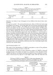
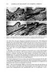
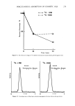
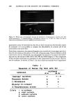
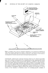
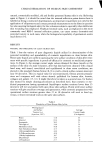
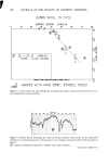
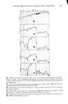
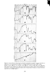
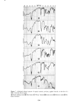
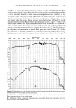
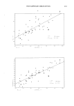
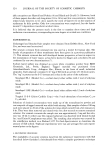
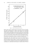
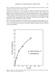
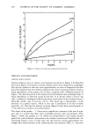
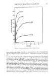
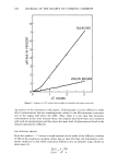
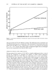
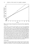
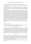
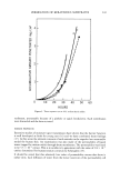
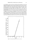
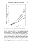
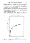
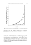
CHARACTERIZATION OF HUMAN SKIN CHEMISTRY 303 therefore, to assess the relative adhesive qualities of such wound fluid films. These qualities must include compatibility with the subjacent cells, supporting their tendency to move smoothly beneath it rather than becoming adherent and resistant to such movement. Table 2 provides contact angle data that characterizes the wettability of a typical proteinaceous film formed by the serous exudate from a "glistening" wound of the forearm skin, the wound having already been characterized in Figure 9, and the exudate characterized in Figure 10 by spectroscopic criteria. Figure 11 plots these contact angle data to yield a critical surface tension value in the mid 20's dynes/cm, a value often found to characterize "biocompatible" natural and synthetic surfaces and argued to have the minimum adhesive potential for living cells (17, 18). The history of the collection and examination of wound fluids goes back over 20 years (19, 20), but the collection of adequate quantities for analysis of the proteins, glycoproteins and lipids present in such fluids has required the use of experimental animals and surgically I FREQUENCY (CM-') 4000 3600 3200 2800 2400 2000 1800 1600 1400 1200 1000 800 650 100 90 80 •7ø 1 •6o z _•$o •4o •3o 2O 10 0 100 90 - • I ................... Figure 12. Demonstration of the different baselines obtained when an internal reflection prism is mounted in different mirror devices for spectroscopic examination. (•) Germanium prism (50 mm x 20 mm x 2 mm) baseline obtained in vertical mirror mounting unit. Note flatter response over most of the spectral range. (•) Same prism baseline obtained in horizontal mirror unit, which allows more convenient and even- pressured skin or liquid cosmetic contact.

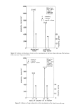
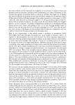
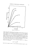
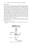
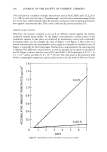
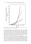
304 JOURNAL OF THE SOCIETY OF COSMETIC CHEMISTS implanted devices. The invasive techniques for placement and removal of collecting receptacles have serious drawbacks when the applicability of the results to mild skin wounds, producing tiny amounts of exudates, are to be considered. The internal reflec- tion, infrared spectroscopic method presented here, supplemented by contact angle measurements of surface properties, and further supplemented by measurements of ellipsometric and electronic parameters for these exudates (! 3, 31), should prove more useful because of the noninvasive character, nondestructive analysis and exceptional sensitivity to thin films of this method. DISCUSSION There is an important departure in the internal reflection, infrared method described here from the normal practice of this technique in other analytical fields. This is the utilization of internal reflection elements in a horizontal position to allow comfortable, gravitationally aided rather than hindered, skin analysis. Horizontal prisms also allow the fascile analysis of fluids, creams and/or medicinal preparations, which would drain from vertical surfaces. This departure from standard methods is not without some sacrifice. Figure 12 demonstrates, for example, the different baselines obtained for the same internal reflection prism mounted in a standard vertical mirror unit vs. the hori- zontal mirror unit preferred for cosmetic and wound-healing studies. The "baseline" quality for the simpler vertical mounting unit is considerably flatter and spectroscopi- cally more desirable than that for the horizontal device. The extreme, concave down, skewing of the baseline for the horizontal device is a disadvantage that must be ac- cepted in order to avoid more serious difficulties experienced when a volunteer is asked to hold a skin surface, with constant pressure, against the vertical face of an internal reflection prism or when drainage causes redistribution of a cosmetic prepara- tion on the prism face during analysis. Nevertheless, there are numerous circumstances in which the simpler setup and supe- rior baseline of the more standard internal reflection, spectroscopic devices may be ac- commodated to the needs of cosmetic or dermatologic analyses. As an example, the rapid analysis of wound fluid components collected in the crevice between shear- separated epidermal layers of friction blisters may be accomplished by first evaporating the fluid to dryness on a horizontal prism face and then turning the prism 90 ø for mounting in a standard vertical unit. Figure 13 presents an internal reflection infrared spectrum of the essentially pure proteinaceous components present in the fluid beneath such a friction blister on human skin and compares it with the spectral baseline for the clean prism in a vertical mounting device. Although friction blisters are very common causes of civilian and military disabilities, they have received very little scientific attention (22). Perhaps the availability of the simple analytical technique described here will allow their more careful examination and the development of improved wound dressings for their more rapid healing. Finally it must be noted that as cosmetic and medicinal preparations begin to incorpo- rate more natural products, and especially products of proteinaceous origin (23), the differentiation among natural skin, damaged skin and the proteinaceous ingredients of various cosmetics, salves, or ointments will become a great deal more difficult than has been the evaluation of cosmetic or therapeutic agents of predominantly hydrocarbon composition. In the former cases, it will be necessary to resort to refinements in the
Purchased for the exclusive use of nofirst nolast (unknown)
From: SCC Media Library & Resource Center (library.scconline.org)