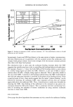
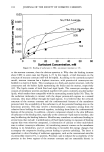
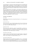
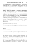
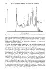
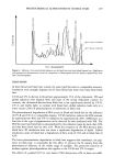
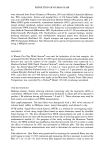
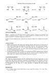
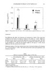
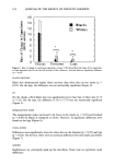
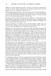
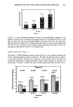
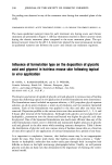
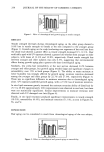
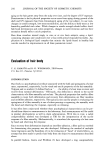
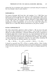
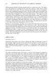
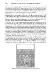
PHOTOCHEMICAL ALTERATIONS IN HUMAN HAIR 203 24 h on a laboratory shaker at 150 rpm. The liquid was filtered off, and the residue was extracted twice with 50 mL each of the solvent mixture as described above. The combined lipid extracts were reduced in volume on a rotary evaporator at 30øC, taken up in 3 ml n-hexane/ispropanol 3:2 (v:v), and stored at -20øC. THIN-LAYER CHROMATOGRAPHY Before TLC separation of the lipid extracts, 10 X 20-cm HPTLC-nano-plates (Mach- erey-Nagel) were washed and activated at 105øC for 30 min. The extracts were applied with an automatic spotting device by Camag. For quantitative determination of the cholesterol fraction, TLC plates were developed subsequently in three solvent systems. The solvent systems were: 1. heptane/ethyl acetate (50:50, v:v) to a running distance of 7.5 cm 2. chloroform/methanol/water (65:36.5:2.5, v:v:v) to a running distance of 8.5 cm and 3. diethyl ether/hexane/acetic acid (50:48.5:2.25, v:v:v) to a running distance of 9.75 cm. For quantitative determination of the fatty acid fraction, the extracts were diluted 1:10. The TLC plates were developed subsequently in two solvent systems. The solvent systems were diethyl ether/hexane/acetic acid (50:48.5:2.25, v:v:v) to a running dis- tance of 6.5 cm and diethyl ether/hexane (3:97, v:v) to a running distance of 9.75 cm. After development, the TLC plates were charred with 10% cupric sulfate in 8% phos- phoric acid (22). DENSITOMETRIC DETERMINATION OF THE LIPID AMOUNT The quantitative determination of the charred cholesterol and fatty acid fractions was carried out with a Hirschmann Elscript 400 © densitometer at 546-nm remission. RESULTS IRRADIATION OF HAIR SAMPLES The black and blond hair samples were irradiated for a period of 6 weeks (1008 h) with UV-B, UV-A, visible light, IR, or global radiation as previously described (12). IL FROM UNIRRADIATED BLOND AND BLACK HAiR SAMPLES The lipid fractions from human hair separated by thin-layer chromatography were rendered visible by charring and identified by using reference substances (REF). The resulting lipid patterns show the influence of specific irradiation ranges on the IL from blond hair samples (Figure 1) and on the IL from black hair (Figure 2). According to the literature, the main fractions of IL from human hair are squalene, cholesterol ester, free fatty acids (FFA), cholesterol, polar lipids, and some fractions that have not been identified up to now (cf. Figures 1 and 2: unirradiated sample) (1,23). Based on results obtained in wool research, it is assumed that the spots between

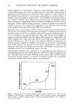
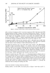
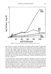
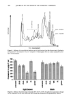
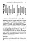
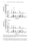
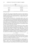
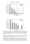
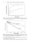
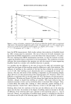
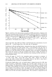
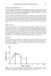
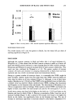
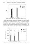
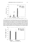
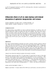
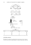
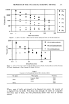
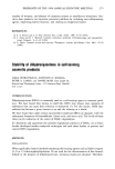
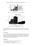
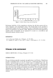
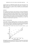
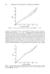
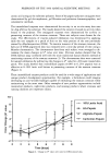
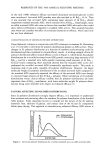
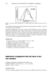
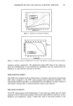
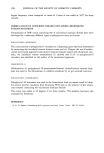
204 JOURNAL OF THE SOCIETY OF COSMETIC CHEMISTS I I I u_ 4-. o I_l_l C n,,' zD squalene cholesfero[ esfer free faffy acids cho[esfero[ nor idenfified polar [ipids Figure 1. Lipid pattern following thin-layer chromatography of IL from unirradiated (untreated) and irradiated [with specific ranges of sunlight (global, IR, VIS, UV-A, UV-B)] blond human hair. REF = reference mixture. cholesterol and the polar lipids consist of several cholesterol oxidation products and that their content can serve as an indicator of oxidative damage of keratin fibers (21). In unirradiated blond and black hair the lipid patterns and intensities of these spots are about the same, which indicates that the qualitative and quantitative composition of the IL from both hair types is independent from the type of pigmentation. In order to obtain a better quantitative estimation in the following, the densitograms of some lipid pat- terns are compared. In these the extinction is a measure of the charring intensity and thus of the amount of a particular lipid fraction. In Figure 3 the lipid patterns from blond and black human hair are compared. The above-mentioned main fractions of the IL are clearly separated from each other in peaks with high extinction. The amounts of polar lipids, cholesterol, cholesterol ester, and squalene are approximately the same in both hair types. Black hair yields a higher peak for the fatty acids and smaller peaks in the area of unidentified lipids. This comparison shows that the composition and amount of IL from blond and black hair largely corre- spond to each other. However, the apparently lower amount of FFA and the larger proportion of unidentified peaks in the area of lipid oxidation products may indicate that blond hair is more susceptible to oxidation than black hair. INFLUENCE OF GLOBAL RADIATION ON LIVID FRACTIONS FROM BLOND AND BLACK HAIR The influence of global radiation on the lipid composition of blond and black human
Purchased for the exclusive use of nofirst nolast (unknown)
From: SCC Media Library & Resource Center (library.scconline.org)