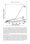
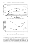
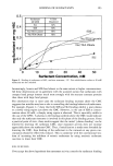
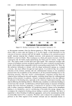
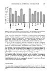
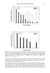
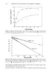
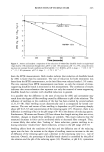
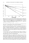
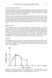
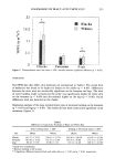
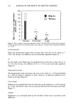
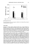
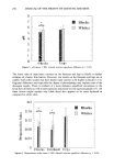
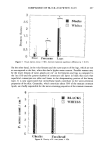
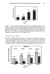
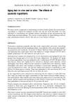
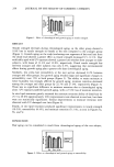
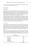
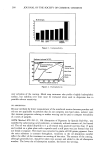
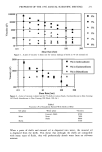
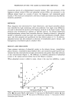
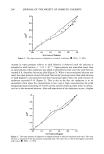
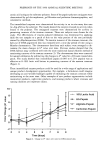
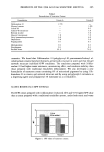
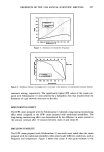
238 JOURNAL OF THE SOCIETY OF COSMETIC CHEMISTS Differences in lipid composition may lead to variations in the amount of bound water (19,20). It was also recently reported that the ceramide content of black skin was significantly lower in comparison to white skin (21). Results regarding hydration levels in blacks and whites from the study by Berardesca et al. (10) closely parallel results from our study. Berardesca et al. found that the water content on the volar forearms was higher for white skin, whereas it was lower for black skin on the dorsal (sun-exposed) aspects of the forearms. The density and size of hair follicles may also lead to differences in skin hydration (18). Racial differences might also be attributed to the melanin content, the packaging of the melanocytes, and their ability to prevent the pronounced epidermal photodamage that occurs in light-skinned individuals (22-24). Though the hydration level in blacks varied in the sites evaluated, TEWL seems to be lower in blacks, irrespective of the water content and sun-induced damage. This sup- ports the notion that blacks possess a superior horny layer barrier. Berardesca et al. (10) found that elastic recovery was significantly higher for whites than for blacks on the volar forearm but significantly lower for whites than for blacks on the dorsal forearm, probably reflecting greater actinic damage in the latter site. In our study, elastic recovery was higher on the cheeks (p 0.05) and legs for black skin as compared to white skin (Figure 4). Biomechanical properties of the skin depend to a large extent on the collagen and elastic components of the dermis (25). According to Montagna et al., black skin has fewer elastic fibers but has greater numbers of oxytalan and elaunin fibers (17). On the contrary, it has also been stated that black skin has more elastic fibers than white skin (26). The higher water content of black SC may also be responsible for the higher elastic deformation observed on the cheeks. We found that pH was significantly lower in blacks on the cheeks (p 0.05) and directionally lower on the legs (Figure 5). A better skin barrier may contribute to the lower skin pH in Blacks. Lactic acid and dicarboxylic amino acids in sweat secretions mixed with sebum may influence skin pH. It has also been postulated that though sweat decreases skin surface acidity, evaporation of sweat causes acidity to increase (27). Since the number of sweat glands in blacks has been found to be higher (17), it is probable that higher amounts of the water-soluble components, which contribute to skin surface acidity, may be left behind on the skin after evaporation of sweat. It has been reported that the activity and size of sebaceous glands are greater in blacks than in whites (28). Hydrolysis of fatty acids, which form a part of the sebaceous lipids, may also contribute to the lower pH in black skin. The desquamation index was found to be significantly lower for blacks than for whites on the cheeks and forehead. It was only slightly lower for blacks as compared to whites on the legs (Figure 6). This may reflect the moisturizing properties of sebum. Analysis of visual grading of dryness showed that it was significantly higher for blacks than for whites on the legs and slightly lower for blacks as compared to whites on the face (Figure 7). There seems to be some disagreement between visual grading scores on the face and legs, which could be fiecause visual grading may be unreliable and is influenced by ambient conditions (13,29). Besides, the high dryness scores (visual) for blacks may be because scales are more perceptible on black skin rather than on white skin. We did not find differences in the density of aerobes between blacks and whites. Though

COMPARISON OF BLACK AND WHITE SKIN 239 no significant differences in the density of P. acnes were found between the two races, there appears to be a trend, with black skin showing a higher P. acnes count. This may be due to the increased sebum output in blacks (28). CONCLUSIONS This study found statistically significant differences in TEWL, electrical capacitance, skin pH, and elasticity between black and white skin. The TEWL, continuous capac- itance, and skin pH data support the hypothesis that black skin has a better barrier function than white skin. The capacitance and elasticity data indicate that black facial skin is more hydrated and elastic than white facial skin. Findings from this study may lead to a better understanding of cosmetic requirements for black skin. REFERENCES (1) M. L. Thomson, Relative efficiency of pigment and horny layer thickness in protecting the skin of Europeans and Africans against solar ultraviolet radiation, J. Physiol., 127, 236-246 (1955). (2) D. A. Weigand, C. Haygood, and J. R. Gaylor, Cell layers and density of Negro and Caucasian stratum corneum, J Invest Dermatol., 62, 563-568 (1974). (3) R. P. Rienertson and V. R. Wheatly, Studies on the chemical composition of human epidermal lipids, Jo Invest. Dermatol., 32, 49-59 (1959). (4) E. K. Marshall, V. Lynch, and H. W. Smith, Variations in susceptibility of the skin to dichloro- ethylsulfide, J. Pharmacol. Exp. Ther, 12, 291-301 (1919). (5) D. A. Weigand and M. M. Mershon, The cutaneous irritant reaction to agent o-chlorobenzylidene malononitrile (CS): Quantification and racial influence in human subjects, Edgewood Arsenal Technical Report 4332 (1970). (6) E. Berardesca and H. I. Maibach, Sensitive and ethnic skin: A need for special skin-care agents? Dermatol. Clin., 9, 89-92 (1991). (7) E. Berardesca and H. I. Maibach, Racial differences in sodium lauryl sulfate induced cutaneous reaction: Black and white, Contact Dermatitis, 18, 65-70 (1988). (8) L. C. Johnson and N. L. Corah, Racial differences in skin resistance, Science, 139, 766-777 (1976). (9) C. L. Janes, J. Worland, and J. A. Stern, Skin potential and vasomotor responsiveness of black and white children, Psychophysiology, 13, 523-527 (1976). (10) E. Berardesca, J. Rigal, J. L. Leveque, and H. I. Maibach, In vivo biophysical characterization of skin physiological differences in races, Dermatologica, 182, 89-93 (1991). (11) S. W. Babulak, L. Wo Rhein, D. D. Scala, F. A. Simion, and G. L. Grove, Quantification of erythema in a soap chamber test using the Minolta Chroma (reflectance) meter: Comparison of instrumental results with visual assessments, J. Soc. Cosmet. Chem., 37, 475-479 (1986). (12) R. R. Wickett, V. Nath, R. Tanaka, and S. B. Hoath, Use of continuous electrical capacitance and transepidermal water loss measurements for assessing barrier function in neonatal rat skin, Skin Pharmacol., 8, 179-185 (1995). (13) P. Williamson and A.M. Kligman, A new method for the quantitative investigation of cutaneous bacteria, J. Invest. Dermatol., 45, 498-503 (1986). (14) H. Schatz, A.M. Kligman, S. Manning, and T. Stoudemayer, Quantification of dry (xerotic) skin by image analysis of scales removed by adhesive discs (D-squames), J. Soc. Cosmet. Chem., 44, 53-63 (1993). (15) H. I. Maibach, Racial and skin color differences in skin sensitivity: Implications for skin care prod- ucts, Cosmet. Toiletr., 105, 35-36 (1990). (16) F. Kompaore, J. P. Marty, and Ch. Dupont, In vivo evaluation of the stratum corneum barrier function in blacks, Caucasians and Asians with two noninvasive methods, Skin Pharmacol., 6, 200-207 (1993). (17) W. Montagna and K. Carlisle, The architecture of black and white skin, J. Am. Acad. Dermatol., 24, 929-937 (1991).
Purchased for the exclusive use of nofirst nolast (unknown)
From: SCC Media Library & Resource Center (library.scconline.org)