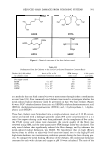
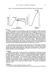
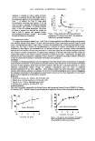
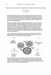
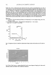
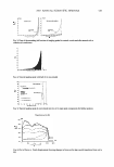
2007 ANNUAL SCIENTIFIC SEMINAR lipophilic (tocopherols) as well as hydrophilic (ascorbate, urate, GSH) antioxidants was detected upon 0 3 exposure and it was accompanied by a rise in lipid peroxidation measured as 4- hydroxylnonenal (4-HNE) content using both Western blot and immunohistochemical analysis. 589 The further step was to investigate whether the antioxidant depletion and the increase of skin lipid peroxidation products can lead to a cellular active responses and to a modulation of skin pathopshysiology. When hairless mice were exposed for 6 days to 0·8 ppm for 6 h day and the homogenized whole skin was analysed, the increase of proinflammatory markers such as cyclooxygenase-2 (COX-2), nitric oxide synthase (iNOS) and metalloproteinases (MMP's) expression was detected together with markers of cellular stress such as heat shock protein (HSP)32, also known as hame oxygenase-1 (H0-1), HSP 27 and HSP 70. It is therefore possible that bioactive compounds generated by products of 03 exposure may be responsible for the induction a stress insult and the release from the skin tissue of inflammatory markers that can than modulate skin inflammation, skin ageing and wrinkles formation. 03 is also able to modulate skin mouse differentiation and the proliferation measure as increase of proliferating cellular nuclear antigen (PCNA) which is a protein identified as the polymerase associated protein synthesized in the early G1 and S phases of the cell cycle involved in DNA replication and repair and keratin 10 (KlO) which is a protein produced in well differentiated, suprabasal keratinocytes It is not clear how 03 displays its effects, but recent studies have shown that it is able to induce the activation of the transcription factor, NF-KB, by phosphorylation of the kinase, IXBa Finally, in our more recent work on wound-healing we were able to also show that when aged animals (18 months) were exposed to 03 the rate of wound closure was significantly delayed when compared to the young animals. Collectively, our data demonstrate that skin exposure to 03 not only affects antioxidant levels and oxidation markers in the outermost stratum corneum layer, but also induces cellular stress responses in the deeper cellular layers of the skin and this can alter skin physiology.

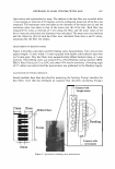
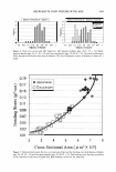
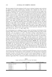
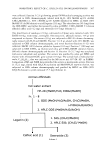
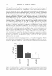
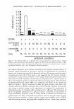
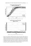
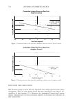
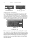
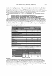
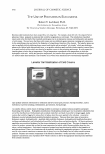
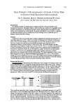
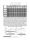
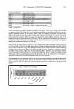
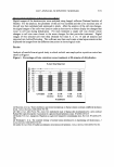
590 JOURNAL OF COSMETIC SCIENCE MODULATING MC 1 R ACTIVITY THROUGH THE USE OF BIOMIMETIC PEPTIDES Kristen Potts, P. Dow, S. Kautz, C. Murphy and C. Zorzopian Active Concepts, LLC Melanogenesis, or the production of melanin for skin pigmentation, begins with the assembly of small chemical messengers in the upper layers of the skin. Lighter skin tends to have lower basal levels of melanogenesis, but exposure to UV radiation or other environmental stress generally induces the amplification of melanin production. One such chemical messenger is Alpha-Melanocyte Stimulating Hormone, or a.-MSH. Melanocyte stimulating hormones belong to a group called the melanocortins, which includes Adrenocorticotropic Hormone (ACTH), a.-MSH, �-MSH, and y-MSH. These peptides are all excision products of a large protein called pro-opiomelanocortin (POMC), but a.-MSH provides the most significant peptide activity. The a-MSH then migrates to and stimulates the melanocortin receptor MCIR, and the resulting cascade of biochemical processes yields an increase in melanin production. Though a.-MSH is an agonist at the Melanocortin-1 Receptor (MClR), there also exist antagonists, such as Agouti Signal Protein (ASP). ASP is able to successfully bind to the MCIR receptor and block the production of a-MSH-stimulated eumelanin (dark melanin), while still allowing the production of pheomelanin (light melanin). In this experiment, novel analogs of a.-MSH and ASP have been isolated. These analogs have key amino acid sequences that were intended to induce changes in skin pigmentation through interactions with the Melanocortin-1 Receptor. To verify this, the analogs and liposomal compositions thereof were tested using a standard MatTek MelanoDerm Assay. This skin model consists of normal, human-derived epidermal keratinocytes and melanocytes that have been co cultured to form a multilayered, highly differentiated model of the human epidermis. The Melanoderm tissues are cultured on specially prepared cell culture inserts using serum free medium. Under appropriate conditions, the melanocytes within this model undergo melanogenesis, leading to melanin accumulation within the tissues over time, which can be influenced by test materials that can either increase (skin darkening agents) or decrease (skin lightening agents) melanin synthesis. With this model, the water-soluble test materials will be directly applied to the surface of the Melanoderm-tissue. Test materials were incubated with the Melanoderm tissue for 14 days. During this period, the tissues were analyzed as follows: (1) biochemical assays for melanin content [day 14], (2) observation of tissue darkening/lightening [days 0, 7, 14], and (3) MTT analysis [day 14]. A MTT assay is a colorimetric analysis of the metabolic activity of the cell, which is a reflection of cell viability. Reduction in MTT by mitochondria results in the formation of insoluble purple formazin crystals that are extracted from the cells with isopropanol and quantified spectrophtometrically. The intensity of the purple color is directly proportional to the metabolic activity of the cells and inversely proportional to the toxicity of the test material. Samples of the two biomimetic peptides were isolated and prepared without preservatives as shown in Table 1. The liposomal dispersions have particle sizes below 250nm. The test materials were prepared by Active Concepts, LLC and shipped to Biolnnovation Laboratories for the assay.
Purchased for the exclusive use of nofirst nolast (unknown)
From: SCC Media Library & Resource Center (library.scconline.org)