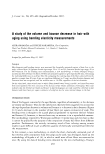
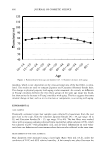
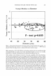
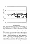
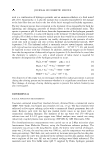
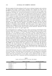
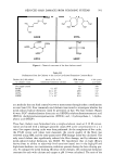
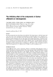
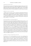
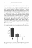
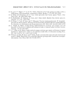
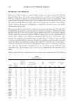
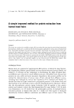
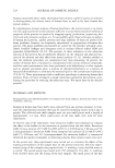
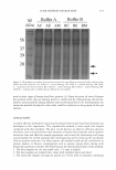
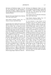
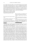
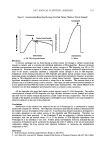
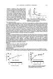
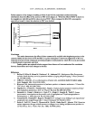
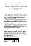
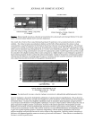
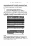
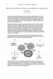
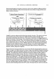
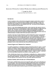
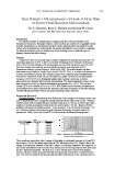
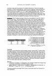
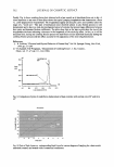
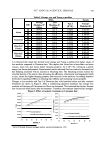
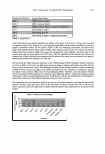
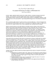
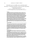
2007 ANNUAL SCIENTIFIC SEMINAR 555 octanoate is required to reach a phase inversion. Acosta et al. proposed that this effect might be due to the preferential partition of the hydrophilic linker in water 11 1 • In addition, the Type IV single phase microemulsion containing equal volumes of oil and water in Figure 1 required the following minimum concentrations: 2.8% lecithin, 3% sodium octanoate (ratio ~l) and 8.4% sorbitan monooleate. The system of Figure 1 requires less than half the lecithin (to form a Type IV system) than standard lecithin polyethyleneglycol-ethanol systems141, and avoided the need for alcohol as cosurfactant In vitro permeation studies 4.5 7 4.0 j .l 3.5 1 3S 3.0 .\ a;. 2.5 ·j , Type I j 2.0 ·! .i 1.5 ·j "'.:_�� Type Ill .J 1.0 1 Typell • --...-� .,, -L o.5 -l - - - ·=--·-··-··-·-··• 0.0 •:••••••••• ....... T .. •••••••••••••• • ••••••• .. •••••••,•••..•••••.. •••••r•••• .. •• ......... 0.0 1.0 2.0 3.0 4.0 5.0 SO/le Figure 1. Phase map for the linker microemulsions prepared at 22°C and 0. 9%w/w NaCL The dotted line indicates the optimwn formulation. To conduct the pem1eation studies Type I and Type II systems produced with different lecithin concentrations were carefully selected (using Figure 1) in such a way that micelles (Type I) and reverse micelles (Type II) would retain a constant diameter (about 10 nm). Figure 2, presents the transdermal flux of lidocaine formulated in Type I (Figure 2A) and Type II (Figure 2B) microemulsions as a function of lecithin concentration in the system. According to these figures, the transderrnal flux of lidocaine increases with increasing lecithin concentration between 0% to 4% lecithin. We have also noted that it is within this region that the lidocaine absorption increases with increase in lecithin concentration. A Langmuir-type adsorption of micelles and reverse micelles (within the internal lipid surface of the stratum comeun) is proposed to explain these results. Additionally, the higher flux obtained in Type II microemulsions (compared to Type I systems) is explained by the higher solubility of lidocaine in Type II systems and its tendency to associated with surface active molecules. Conclusion In summary, the adsorption/permeation of active ingredients from linker based lecithin microemulsions is dependent on the lecithin concentration. As the lecithin concentration increases, more micelles (or reverse micelles) are formed and more of these aggregates absorb into the stratum comeum. After the skin is saturated (achieved at a certain surfactant concentration), further increase in surfactant concentration does not result in improved absorption/pemieation of the active ingredient These findings suggest that future formulation studies should consider optimizing the surfactant concentration for the level of topicaVtransdermal delivery required. Minimizing the surfactant concentration in these applications would result in lower cost, but more importantly in lower risk of allergic reaction triggered by the formulation excipients. References 1. Acosta et al., Environ. Sci. Technol., 39, 1275-1282, 2005. 2. Sabatini et al., Colloids Interface Sci., 8, 316-326, 2003. 3. Yuan et al. Int. J. Pharm. Submitted Januacy 2007. 4. Corswant et al., Langmuir, 14, 6864-6870, 1998. Acknowledgements This work was partially supported by the Natural Science and Engineering Research Council (NSERC) of Canada. We thank the SCC- Ontario Chapter for providing financial support for Jessica Yuan's participation at this meeting. �m A E �m °' ::::J \,,.,/ X ::::J LL 0 Type I (micelle) 2 3 Lecithin,% w/w 4 � E u .c ........ °' ::::J \,,.,/ X ::::J u:: 0 Type II (reverse micelle) 2 3 4 Lecithin,% w/w Figure 2. Transctermal 11ux of Hdocaine 1btmula1Bd in Type I (PartA) and Type II (PartB) microem.Jlsions cmtaining differentledthin ooncentrations.

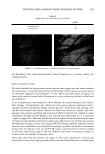
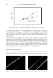
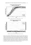
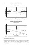
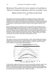
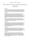
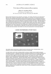
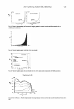
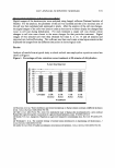
556 JOURNAL OF COSMETIC SCIENCE RHEOMORPHOLOGICAL CHANGES IN MASCARA TEXTURE RELATED TO f ILLING STRESS Yelena Loginova, Ralph Macchio and Alan Farer Coty International Research and Development Center 410 American Road) Morris Plains) NJ 07950) USA Abstract The performance of mascara has a strict correlation with its rheological properties. As a heavy emulsion, mascara exhibits a complex rheological behavior. The aim of this study was to investigate the shear induced structural changes in mascara formulation related to filling stress. The corresponding microscopic images that show the deformation and recovery of the formula make a valuable contribution to product development. The study attempts to characterize the rheomorphological changes in mascara texture associated with filling. The corresponding images of product performance on false lashes made of human hair illustrate the influence of rheomorphological changes on makeup results. Materials and Methods: Rheomorphological data were obtained with a Brookfield rotational speed and stress controlled R/S - CPS Rheometer with a plate measuring system (plate - C25-2), using software 2.6. Visualization of images was obtained with an Olympus BH2 microscope with a digital camera. Total image magnification with a 17" screen was 800X. Mascara samples were filled manually according to a routine lab procedure and with a GRISO NA MA pump filling machine. The mascara bulk was studied before the filling. The samples were evaluated immediately after the filling, then 24 hrs and 72 hrs later. Results were generated using the standard rheological test methods: viscosity measurements with controlled stress and controlled shear rates, yield stress measurements, thixotropy and recovery. The yield value was obtained by extrapolating to zero on the shear stress-shear rate curve. At the end of the recovery test the measured material was collected for microscopic evaluation. The mascara formulation having of7% of polymer, 28/50 w/w percents of water/oil ratio and 10% of a pigment phase containing of the Black Iron Oxide was employed for this study. Results and discussion We found similar rheological behavior of the formula after the hand filling through syringe and filling machine. Fi gu re 1 shows the compression of the formula recovery before and after the filling.
Purchased for the exclusive use of nofirst nolast (unknown)
From: SCC Media Library & Resource Center (library.scconline.org)