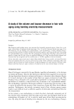
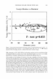
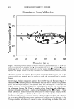
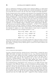
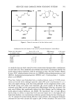
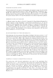
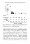
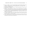
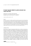
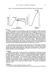
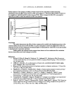
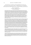
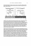
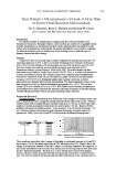
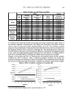
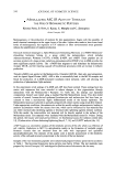
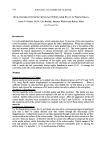
2007 ANNUAL SCIENTIFIC SEMINAR 595 Measurement of Changes in Keratinocyte Cell Size Digital images of the keratinocytes were analyzed using ImageJ software (National Institute of Health). For the analysis, the perimeter of the cell was outlined and the cross sectional area of the cell was then calculated and expressed in pixels. After the analysis of the cell size changes, the digital images of the cells were used to create a short movie to better display the changes that occur in cell size during dehydration. For each treatment a single cell was chosen whose changes in cell size were closest to the mean changes for that particular treatment. Digital images of this selected cell were then obtained for times 0, 5� 10, 15 and 20 minutes and imported into Adobe Photoshop. This software was then used create a time lapse animation that displayed the images from the different time points in chronological order. Results Analysis of results from a typical study in which sorbitol was employed as a positive control are shown in Figure 1. Figure I. Percentage cell size retention verses treatment at 20 minutes of dehydration % Cell Size Retention C :8 100 C 90 +-----------t---------=�----+------i JI 80 --+-----+----1---- 1 70 • 60 ii 50 ii 40 U 30 1: 20 � 10 l. a Untreated 0.5% Sorbitol 0.5% Active Treatment 1.0% Active 2.0% Active [1] Bouwstra JA et al., Water distribution and related morphology in 1mrnan stratum comeum at different hydration levels. J Invest Dermatol. 120 (2003) 750-758. [2] Chrit L et al., Skin Pharm Phy. An in vivo randomized study of human skin moisturization by a new confocal Raman fiber-optic microprobe: Assessment of a glycerol-based hydration cream. 19 (2006) 207-215 [3] Mariko H.C et al., Aquaporin-3 functions as a glycerol transporter in mammalian skin Biol Cell. 91 (2005) 479- 486. [4] Warskulat U. et al., The osmolyte strategy of normal human keratinocytes in maintaining cell homeostasis. J Invest Dermatol. 123 (2004) 516-521. [5] Janeke Get al., Role of Taurine accumulation in keratinocyyte hydration J Invest dermtol. 121 (2003) 354-361.

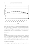
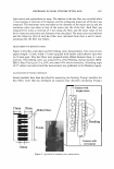
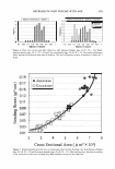
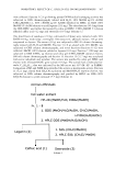
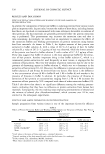
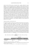
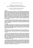
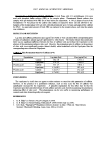
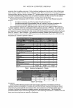
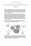
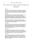
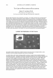
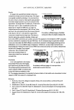
596 JOURNAL OF COSMETIC SCIENCE DOES MELANIN HAVE AN SPF AND CAN IT BE MEASURED? Howard Epstein1'2, Prashiela Manga3, Amu Koshoffer*, David Story2, Tony Simion2, Raymond Boissy* 1 Union Institute and University of Cincinnati, OH 2 Kao Brands Company, Cincinnati, OH -'Department of Dermatology, New York University School of Medicine, NY, NY * Department of Dermatology, University of Cincinnati College of Medicine, Cincinnati, OH Introduction The Food and Drug Administration (FDA) describes Sun Protection Factor Value (SPF) as the UV energy required to produce a minimal erythema dose (l\,1ED) on protected skin divided by the UV energy required to produce an MED on unprotected skin. Limited studies have been performed to determine the specific SPF of melanin. In order to standardize testing, we have begun to develop an assay using methodology initially designed for the determination of the SPF of sunscreen products. One accepted protocol measures SPF value of sunscreen products after application of 2 milligrams per square centimeter of product is applied to skin in vivo and exposed to UV irradiation. The SPF value is the reciprocal of the effective transmission of the product viewed as a UV radiation filter (1). The Optometries SPF-290S was developed an in vitro method to measure SPF values of various types of material containing sunscreen products. The instrument was designedto detect low levels of transmitted light using a high output continuous 125-watt xenon lamp. Light from the lamp passes directly through the sample. The recommended protocol requires a concentration of 2 uL/cm2 of sample is mixed evenly in support medium and spread on a quartz plate. A sheet of Transpore Tape™ (TT) is placed over the quartz plate to simulate human skin. The instrument makes automatic measurements over the entire area integrating any irregularities caused by spreading technique (2) A few investigators have reported that the SPF of in vivo melanin ranges from SPF 2-9.68 (3,4,5). In this study we report on the results of a protocol developed to measure the SPF of melanin extracted from cultured human melanocytes from donors with light to dark skin. Materials and Methods The SPF of melanin was determined using an SPF-290S Analyzer System manufactured by Optometries LLC. Neonatal human and mouse melanoma melanocytes were cultured to confluence in either MCDB153 medium, supplemented with 3% fetal bovine serum, 5µg/ml insulin, 2ug/ml transferin, lµg/ml Vitamin E, 0.6ng /ml bFGF, 13µg/ml bovine pituitary extract, SnM 12-0-tetradecanoylphorbol-13-acetate and 1% penicillin-streptomycin or DMEM supplemented with 8% fetal bovine serum, lmM glutamine, 0.5mM sodium pyruvate and 1% penicillin-streptomycin media, respectively. Cells from multiple culture flasks were combined in a centrifuge tube, pelleted and rinsed twice in Hank's buffered saline solution (HBSS). The melanin pellets were weighed and were either mixed with 0.2N sodium hydroxide and a sunscreen-free moisturizing lotion orl % gelatin in HBSS used as support medium to facilitate spreading on TT that was attached to the quartz test plate. Melanin combined with gelatin or lotion was applied per Optometries protocol (2). Results and Discussion In order to determine the optimal conditions for measuring the SPF of melanin, we evaluated the following conditions on outcome as reproducibility: 1. melanin solubilization, 2. application vehicle and 3. amount of melanin. In the initial studies quantities of melanin ranging from 0.12 grams to 0.30 grams mixed with 110 uL sodium hydroxide did not provide enough material to evenly spread on TT. A quantity of 220 uL gelatin or lotion was determined to be the optimal volume of support medium to facilitate spreading of melanin on TT. It was then necessary to determine the optimal amount of melanin required to obtain consistent readings from the SPF Analyzer. A quantity of 0.30 grams of mouse or human melanin obtained through cell culture was determined to be the optimal amount for this study. SPF baseline reading for 1 % gelatin in HBSS and test lotion was obtained as shown in Tables 1 and 2. SPF data obtained for cultured melanocytes was compared with SPF readings for synthetic stock melanin solution of0.01 grams solubilized in 10ml of0.2N sodium hydroxide (1000 ug/mL). A 1:10 dilution (100 ug/mL) of stock melanin solution was tested for SPF. A quantity of 300 uL (0.30 grams) synthetic melanin at 1000 ug/mL and another sample at 100 ug/mL was measured for SPF as shown in Tables 3 and 4.
Purchased for the exclusive use of nofirst nolast (unknown)
From: SCC Media Library & Resource Center (library.scconline.org)