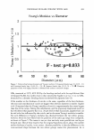
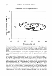
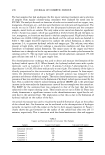
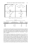
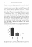
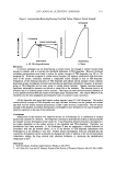
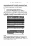
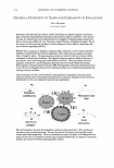
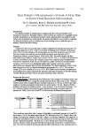
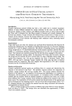
528 JOURNAL OF COSMETIC SCIENCE findings mentioned above imply that human hair proteins could be used as an indicator in distinguishing the diverse status of human hairs as well as for basic human hair protein analysis. In comprehensive protein analyses of human head hairs, the initial concern is to obtain not only quality protein but also amounts sufficient to assay chemical and/or biochemical properties of the proteins in question by mapping/typing, purification, sequencing, hair structural and proteomic analyses, etc. To successfully apply these techniques to analysis of human hair proteins, quality proteins and large amounts of them are needed (10). Some problems, however, are frequently encountered in preparations of human hair protein. The major problems encountered are caused by the presence of highly cross linked disulfide linkages and detergents such as sodium dodecyl sulfate (SDS) and guanidine hydrochloride (11, 12). The presence of these substances in the extraction solution of hair protein has an inhibitory influence on the chemical and physical reac tions of the solution and thus affects hair protein analysis ( 11, 12). Another problem is that the isolation procedures are complicated and time-consuming. In general, the surface of human hairs is lipidized or contaminated with various chemical compounds, and thus some pretreatments have been performed with delipidizing or other reagents such as ethanol, petroleum ether, a mixture of chloroform/methanol, or a mixture of hexane/dichloromethane to eliminate the external lipid or the contaminated substances (7 ,8,10,11). These pretreatments lead to inefficient procedures in extracting human hair proteins. Thus, we have developed a simple extraction method for hair protein, accel erating the procedure by reducing the extraction steps. We report here on the detailed protocol. MATERIALS AND METHODS PREPARATIONS AND PRETREATMENTS OF HUMAN HEAD HAIR SAMPLES, BUFFER SOLUTIONS, AND TEMPORAL ANALYSIS Samples of human head hair shafts were collected from one woman volunteer to mini mize the experimental variations that may be caused by sampling sources such as age or gender. The collected hair shaft samples were cut into small pieces with a length of approximately 1-2 mm. These small pieces of the hair shafts were used for these experiments. In the first step of the experiment, three extraction buffers were employed to compare the effect of the buffers on the extraction of proteins from the hair shafts. Extraction buffer A was a solution of 25 mM Tris-HCl (pH 8.5), 2.6 M thiourea, 5 M urea, and 5% 2-mercaptoethanol. Extraction buffer B was comprised of a solution of 25 mM Tris-HCl (pH 9.5), 8 M urea, and 5% 2-mercaptoethanol. For extraction buffer C, a commercial extraction kit applied commonly in extracting proteins from various tissues was used. In the second step, three experiments were carried out to scrutinize the effects of the washing pretreatments of the sample hairs on the protein extraction. The first experi mental group of hair samples was prepared with 3 x washings, using distilled water as a control. The second group was treated with 75% ethanol for 30 s after 3x washings with distilled water and the drying of the hair samples. The small pieces of the hair

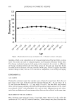
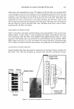
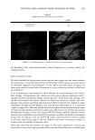
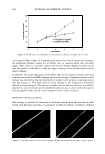
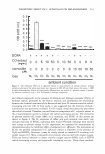
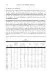
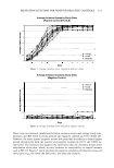
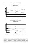
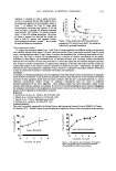
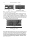
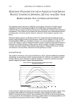
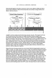
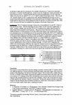
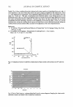
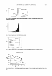
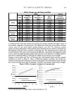
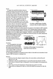
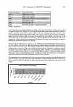
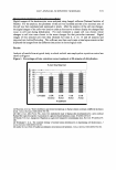
HAIR PROTEIN EXTRACTION 529 samples belonging to the third group were immersed in a mixture of chloroform/ methanol (2:1, v/v) for 24 hr at room temperature after ethanol treatment for 30 s and drying of the samples. In the third step, the effect of the sample preparations of the hair shafts on the protein extraction was compared by preparing two groups of the hair materials. The first group of the hair materials was pulverized in a mortar with a pestle using liquid nitrogen, and then the powdered hair materials were used for protein extraction in the buffer solutions. Another group of the hair materials was directly immersed in the buffer solutions without grinding in the mortar. In the fourth step, various incubation times were introduced to establish the optimal incubation time in extracting the protein from the hair shaft materials. For this experi ment, the hair materials were washed three times with distilled water and incubated at 50°C for 12 different incubation times ranging from 2.5 hr to 50 hr (2.5, 5.0, 7.5, 10, 15, 20, 25, 30, 35, 40, 45, and 50 hr). The extraction mixtures (containing the buffer solution and the hair materials) collected at the given incubation times were filtered through three layers of nylon mesh, and the amounts of protein were quantified to analyze the temporal progress of the protein extraction. EXTRACTION, MEASUREMENT, AND SDS-PAGE ANALYSIS OF HUMAN HAIR PROTEIN Total proteins were extracted by placing the hair materials into a tube or container containing the buffer solutions. After each sample group had been pretreated, 20 mg of the hair materials were incubated in buffer solutions A and B (5 ml per 20 mg materials) at 50°C for the given extraction times. In buffer solution C (a commercial protein extraction kit), the extraction procedures were done according to the manufacturer's manual. After completion of their incubations, the mixtures were then filtered through three layers of nylon mesh (200-mesh size) and the flowthrough was used for the protein measurement. The amounts of protein were measured with the protein-dye binding method of Brad ford (13) using a commercial protein assay kit (Bio-Rad, Hercules, CA). For this analysis, 10 µl of the incubated flowthrough was transferred to a cuvette containing 200 µl of dye solution and 790 µl of distilled water to make 1000 µl in total. The amounts of the total protein secreted into the buffer solutions were then quantitated by detecting the optical density at 595 nm of a wavelength, using a spectrophotometer (HP Agilent 8453, Palo Alto, CA). For analysis of sodium dodecyl sulfate-polyacrylamide gel electrophoresis (SDS-PAGE), 2 ml of the protein flowthrough was concentrated with a Centricon centrifugal filter device (Millipore, Bedford, MA) by centrifuging the devices containing the filtrate at 3000g for approximately 2.5-3 hr at 4°C. Thirty micrograms of the concentrated pro teins were boiled for 5 min and cooled in ice for 2 min, and then analyzed using the Bio-Rad mini-gel system (Bio-Rad, Hercules, CA) according to the manufacturer's manual. A gel with 5 % stacking and 15 % separating acrylamide was employed and visualized after staining with Coomassie brilliant blue G-250. The stained gel was transferred to a container containing a destaining solution (10% isopropanol and 10% acetic acid, v/v) and destained three times with slow shaking.
Purchased for the exclusive use of nofirst nolast (unknown)
From: SCC Media Library & Resource Center (library.scconline.org)