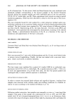
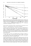
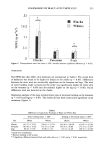
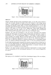
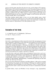
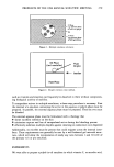
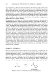
202 JOURNAL OF THE SOCIETY OF COSMETIC CHEMISTS on IL of human hair. To this extent, black and blond human hair were irradiated with simulated sunlight corresponding to the natural sunlight in the Central European latitude during the summer months and with individual parts of the sun spectrum, UV-B, UV-A, visible light, and IR. These experiments were carried out using a special irradiation apparatus, which has been described in detail in the first part of this inves- tigation (12). The non-covalently bound IL were isolated by a mild extraction method under non- oxidizing conditions so as to prevent further reactions of lipids already photochemically oxidized. The lipid extracts were separated by TLC using solvents of different polarity and then charred. Representing all IL, the main fractions--free fatty acids (FFA) and cholesterol--were quantitatively investigated by densitometric scanning of the charred spots. Finally, a photochemical destruction of the IL as a function of the type of irradiation and pigmentation of the hair is discussed. MATERIALS AND METHODS HAIR SAMPLES Untreated black and blond hair was obtained from Herzig Co. as 25-cm-long tresses of European origin. PURIFICATION OF HAIR Hair was extracted for 5 min with dichloromethane and for 30 min with diethyl ether to remove traces of naphthalene. Finally, the hair was washed with a non-ionic deter- gent, rinsed, and stored at ambient humidity. IRRADIATION OF HAIR The hair tresses were irradiated for a period of 6 weeks (1008 h) in individual com- partments with UV-B, UV-A, visible light, IR, or global radiation at RH 70% (12). In each case approximately 10 g of hair were used, the hair was spread parallel and rearranged daily to assure uniform exposure to irradiation. EXTRACTION OF SURFACE LIPIDS In order to remove the external lipids (sebum) and superficial deposits, irradiated hair samples were extracted with n-hexane under non-swelling conditions in a Soxhlet ex- tractor in vacuo according to the method of Schwan et al. (3). EXTRACTION OF INTERNAL LIPIDS Following surface extraction, hair samples were manually cut into ca. 3-mm-long fiber snippets. One gram of this sample was conditioned for 24 h at 20øC and 65% RH, mixed with 50 ml n-hexane/isopropanol/water 6:6:1 (v:v:v) in an Erlenmeyer flask, covered with a layer of nitrogen, protected from light by aluminium foil, and shaken for

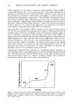
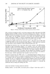
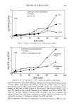
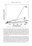
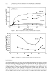
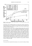
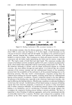
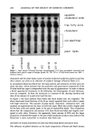
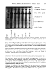
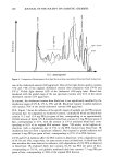
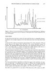
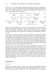
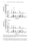
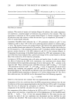
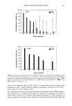
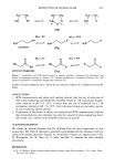
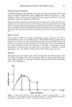
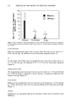
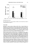
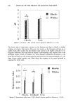
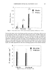
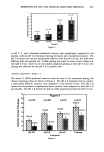
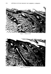
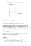
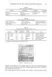
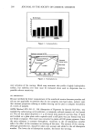
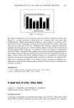
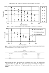
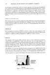
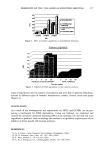
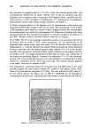
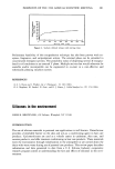
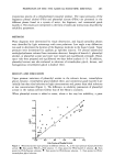
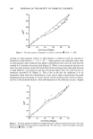
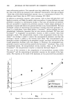
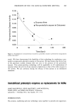
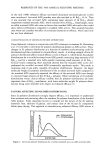
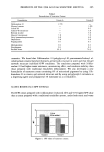
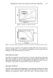
PHOTOCHEMICAL ALTERATIONS IN HUMAN HAIR 203 24 h on a laboratory shaker at 150 rpm. The liquid was filtered off, and the residue was extracted twice with 50 mL each of the solvent mixture as described above. The combined lipid extracts were reduced in volume on a rotary evaporator at 30øC, taken up in 3 ml n-hexane/ispropanol 3:2 (v:v), and stored at -20øC. THIN-LAYER CHROMATOGRAPHY Before TLC separation of the lipid extracts, 10 X 20-cm HPTLC-nano-plates (Mach- erey-Nagel) were washed and activated at 105øC for 30 min. The extracts were applied with an automatic spotting device by Camag. For quantitative determination of the cholesterol fraction, TLC plates were developed subsequently in three solvent systems. The solvent systems were: 1. heptane/ethyl acetate (50:50, v:v) to a running distance of 7.5 cm 2. chloroform/methanol/water (65:36.5:2.5, v:v:v) to a running distance of 8.5 cm and 3. diethyl ether/hexane/acetic acid (50:48.5:2.25, v:v:v) to a running distance of 9.75 cm. For quantitative determination of the fatty acid fraction, the extracts were diluted 1:10. The TLC plates were developed subsequently in two solvent systems. The solvent systems were diethyl ether/hexane/acetic acid (50:48.5:2.25, v:v:v) to a running dis- tance of 6.5 cm and diethyl ether/hexane (3:97, v:v) to a running distance of 9.75 cm. After development, the TLC plates were charred with 10% cupric sulfate in 8% phos- phoric acid (22). DENSITOMETRIC DETERMINATION OF THE LIPID AMOUNT The quantitative determination of the charred cholesterol and fatty acid fractions was carried out with a Hirschmann Elscript 400 © densitometer at 546-nm remission. RESULTS IRRADIATION OF HAIR SAMPLES The black and blond hair samples were irradiated for a period of 6 weeks (1008 h) with UV-B, UV-A, visible light, IR, or global radiation as previously described (12). IL FROM UNIRRADIATED BLOND AND BLACK HAiR SAMPLES The lipid fractions from human hair separated by thin-layer chromatography were rendered visible by charring and identified by using reference substances (REF). The resulting lipid patterns show the influence of specific irradiation ranges on the IL from blond hair samples (Figure 1) and on the IL from black hair (Figure 2). According to the literature, the main fractions of IL from human hair are squalene, cholesterol ester, free fatty acids (FFA), cholesterol, polar lipids, and some fractions that have not been identified up to now (cf. Figures 1 and 2: unirradiated sample) (1,23). Based on results obtained in wool research, it is assumed that the spots between
Purchased for the exclusive use of nofirst nolast (unknown)
From: SCC Media Library & Resource Center (library.scconline.org)