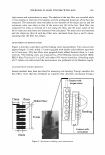
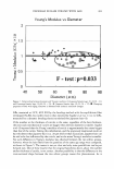
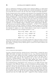
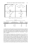
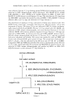
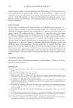
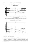
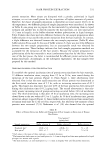
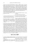
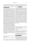
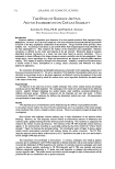
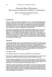
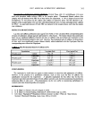
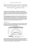
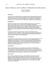
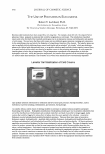
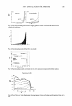
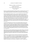
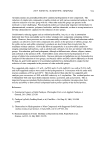
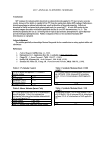
INHIBITORY EFFECT OF C. OFFICINALIS ON MELANOGENESIS 509 0 OH OH caffeic acid ( 1 ) OH HO 0 OGlu loganin (2) OH morroniside acetate ( 4) HO HO 0 OGlu sweroside (3) cornuside (5) Scheme 3 515 nm was monitored. In a similar way, 0.05 ml of solvent (MeOH or water) was added instead of a test sample as a control. The blank was 1.95 ml of MeOH plus 0.05 ml of solvent (MeOH or water). DPPH radical scavenging activity was measured by compari son with the control value. DPPH scavenging rate(%)= [{ABSc -(ABS 5 - ABS B )}/ ABSc] x 100 where ABSc = the absorbance value of the control, ABS5 = the absorbance value of the test sample, and ABSB = the absorbance value of the blank. MEASUREMENT OF CHEMILUMINESCENCE Weak chemiluminescence was observed with compounds from C. officinalis that showed DPPH scavenge activity and EGCG. A half milliliter of cornuside (or caffeic acid) isolated from C. officinalis was mixed with 0.5 ml of 5% (w/w) H2O2 and 0.5 ml of 100 mM KHCO 3 . Using the low-level light detection unit C8801 (Hamamatsu Photonics K.K.), photons from test samples (final cone. 0.05 mM) were measured and integrated for 20 min at room temperature.

510 JOURNAL OF COSMETIC SCIENCE SUPEROXIDE SCAVENGING ACTIVITY Radical production by the reaction with xanthine and xanthine oxidase was detected by a superoxide dismutase (SOD) assay kit, WST (Dojindo Molecular Technologies, Inc.). Briefly, after adding 0.02 ml of compounds from C. officinalis extract to 0.2 ml of WST working solution, each well was mixed. The mixture was incubated at 3 7°C for 20 min. The optical density (OD) at 450 nm was measured as an index of superoxide quantity. INHIBITION OF L-DOPA AUTO-OXIDATION C. officinalis extract was added to 10 µl of 1.4 mg/ml of L-Dopa (Kishida Chemical Co) aqueous solution at room atmosphere to obtain final concentrations of 5, 10, and 50 mg/ml in 50-ml centrifuge tubes. The same volume of 1.4 mg/ml of L-Dopa solution (10 µl) without C. officinalis extract bottled at an ambient atmosphere or under N2 gas in a 50-ml centrifuge tube was used as a control (C. officinalis extract, 0 mg/ml, 02 ambient condition). The absorbances of the mixtures were measured at 405 nm, after being incubated at 3 7°C for one week. MELANIN BIOSYNTHESIS IN CULTURED MELANOMA CELLS B 16 melanoma cells were purchased from Riken Cell Bank. Cells were seeded at 5 x 10⁴ cells/well and cultured for 24 h in minimal essential medium supplemented with 10% fetal bovine serum (Gibco BRL). After changing the medium to fresh medium, com ponents in C. officinalis and arbutin (Merck) dissolved in 30% EtOH where applied, and the cells were incubated for three days. The final concentration of the components in C. officinalis and arbutin was 0.1 mM, and from C. officinalis extract it was 0.025 mg/ml. The same volume of 30% EtOH was used as a control. Cells were harvested by treatment with trypsin, dissolved in 1 ml of 1 N NaOH and 10% dimethyl sulfoxide (DMSO Kishida Chemical Co, Ltd), and then OD at 420 nm was measured as an index of melanin quantity. Cytotoxicity was examined by the MTT method in a colorimetric assay system that measures the reduction of a tetrazolium component into an insoluble forma zan product by the mitochondria of viable cells. After incubation of the cells with the MTT reagent for approximately two to four hours, a detergent solution was added to lyse the cells and solubilize the colored crystals. The samples were read using an ELISA plate reader at a wavelength of 5 70 nm. The amount of color produced was directly propor tional to the number of viable cells. INHIBITION OF DVB-INDUCED PIGMENTATION IN GUINEA PIG Five guinea pigs with brownish pigmentation were used in this experiment. Four separate areas (2 cm x 2 cm) on the back of each animal were irradiated with UVB (250 mJ/cm2 per day) for three consecutive days. On the fourth day through the 14th day, 0.02 ml of arbutin (30 mg/ml), C. officinalis extract (100 mg/ml), or vehicle (50% EtOH) as a control, were topically applied before the daily UVB radiation. The degree of pigmentation was assessed as the delta L * value, that is, the means of L * value differences (before and after the UVB irradiation), with a chromometer (CR-200, Konica Minolta, Inc.).
Purchased for the exclusive use of nofirst nolast (unknown)
From: SCC Media Library & Resource Center (library.scconline.org)