]. Cosmet. Sci., 56, 379-393 (November/December 2005) An examination of non-invasive imaging techniques in the analysis and review of cellulite THERESA CALLAGHAN and KLAUS PETER WILHELM, proDERM Institute for Applied Dermatological Research, Hamburg, Germany. Accepted for publication July 18, 2005. Presented in part at the 23rd IFSCC Congress, Orlando, Florida, October 2004. INTRODUCTION Cellulite, sometimes more specifically referred to as a gynoid lipodystrophy, has recently found itself classified in two new "trendy" ways: aesthetic endocrinology and gender specific medicine. Yet the fact remains unaltered: cellulite is not a skin disorder or disease, nor a tissue dystrophy, but an inevitable reality of the genetic makeup of the female human species wholly interlinked with steroid hormones and external influences. Cellulite is a condition that needs to be managed since it cannot be cured inside the strict confines of cosmetic definition. A clearer understanding of the influences on adipose tissue metabolism, and connective tissue structure, should lead to the develop ment of new test methods to advance a more coherent approach to understanding cellulite. It is unfortunate that as technology continues to advance so does the image of the "perfect body." The "Western" consumer culture latches onto the prevalent self preservation concept of the body, which encourages the individual to adopt strategies to combat aging. Obsession and preoccupation with the body has continued to increase, and Western culture continues to be obsessed with female body size and shape, seeing fatness and thinness as ultimate statements of their worth rather than as descriptions of the ratio of fat body tissue to lean body tissue and body mass index (1). Cellulite affects all women, and up to 30% of their subcutaneous fat can physically appear as visible cellulite. Supermodels, anorexics, and Olympic athletes all have this condition. As a group of individuals they could not be more different, yet they all have one thing in common apart from their gender and connective tissue organization stress. Stress in the skin manifests itself in a number of ways, the most widely acknowl edged being "aging" of connective tissues (2-4). Stress in subcutaneous tissues, i.e., adipose tissue, is less understood (5-7), and the biochemistry of stress in this area of cosmetics has not been evaluated with much consideration. When evaluating the cel lulite condition, be it for a new "active" delivery system or a finished product, devel- 379

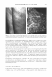
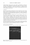
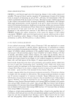
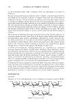
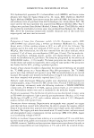
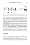
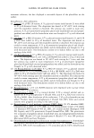
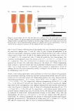
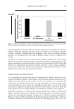
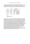
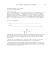
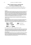
380 JOURNAL OF COSMETIC SCIENCE opment needs to take a wider look at the causes of the condition, not only its manifes tation. There is little coherency or consistency within the scientific literature as to the mani festation of the "cellulite condition" (9-17). From a clinical viewpoint, biochemical markers in adipose tissue metabolism can play a key role in understanding the cellulite condition and, therefore, in determining the effectiveness of an active/product in the management of cellulite. Furthermore, the development of clinical methods, which can correlate in vitro studies, will finally bring coherence and understanding to the scientific literature and to the condition, and will ultimately manage the continued expectations and unmet needs of the consumer. Before examining the available clinical imaging methods for cellulite prognosis and the determination of the effectiveness of cosmetic treatments, an overview of the current understanding (or lack of) of the cellulite condition is required, in terms of adipose structure and influences, and of connective tissue organization. ADIPOSE TISSUE ADIPOCYTE SIZE AND ORGANIZATION It is understood from histological examination that the adipose structure within females is different from that of males. Fat accumulation causes pressure, which results in a "hernia" protruding into the dermis and epidermis (8-10). The observed effect (Figure 1) is of dimpling of the skin, notably on the thighs and buttocks, with histopathological observations clearly highlighting fibrosclerotic tissue structures surrounding the adipose tissue, with resultant striae at the dermal/epidermal interface (8). Adipose tissue is a specialized connective tissue that functions as the major storage site for fat (triglycerides). The major bulk of adipose tissue is a loose association of lipid-filled adipocytes held in a framework of collagen fibers. It also contains stromal, vascular cells including fibroblastic connective tissue cells, leukocytes, macrophages, and pre adipocytes that contribute to structural integrity. Distribution in humans is dependent on genetic and environmental factors. The total and regional masses of adipose tissue are dependent on the number of adipocytes as well as their degree of filling with depot fat. Once new adipocytes are formed, they remain. Increasing numbers of adipocytes have far-reaching consequences for the treatment and prevention of obesity. Currently avail able evidence does not suggest a specific regional regulation of fat cell multiplication in subcutaneous depots, which instead seems to occur to a certain critical degree in the filling of available adipocytes (18). The control of the rate of filling of adipocytes seems to be the main factor in determining the local, regional mass of adipose tissue. The balance between the lipid accumulating and mobilization processes in turn regulates this. The steroid hormones exert major effects on these processes (19,20). It appears likely that the resulting effect of the rate of secretion of various steroid hormones, and the local density of their specific receptors, decides the regional distribution of body fat. GENDER DIFFERENCES Adipose tissue is the source of a critical endocrine signal in the control of body weight.
Purchased for the exclusive use of nofirst nolast (unknown)
From: SCC Media Library & Resource Center (library.scconline.org)