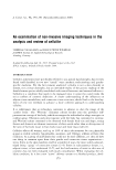
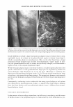
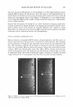
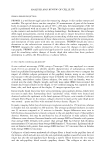
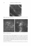
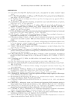
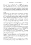
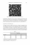
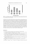
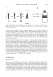
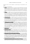
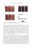
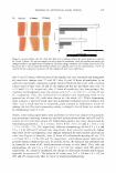
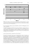
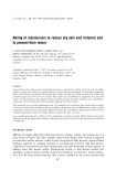
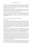
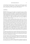
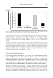
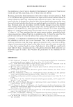
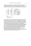
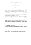
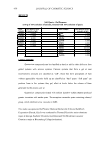
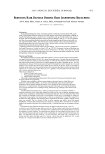
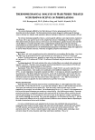
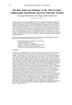
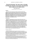
ANALYSIS AND REVIEW OF CELLULITE 381 Figure 1. Clinical aspects of cellulite dimpling (a) of the skin on the female thighs is a visible characteristic of cellulite. Note also the torsion of the skin diagonally across the skin. The full-thickness biopsy (b) shows the fibrosclerotic nature of the connective tissue, with corresponding bulging of the surrounding adipose. Gender differences include a larger subcutaneous adipose tissue in women than in men, explainable in part by a depot in the gluteal-femoral region in women ("pear shape"), which is essentially absent in non-obese men. Men, on the other hand, have a larger proportion of their adipose tissue localized intra-abdominally ("apple" shape). In addi tion, the gluteal-femoral fat cells in women are specifically enlarged, and demonstrate a higher activity of the enzyme lipoprotein lipase. While the larger adipose tissue in non-obese women is considered genetically linked, the specific characteristics of the gluteal-femoral adipocytes are regulated by female sex steroid hormones (20). Hormones are by far the most influential in regulating the deposition of gluteal-femoral adipose tissue (21,22). The sex steroid hormones are never active alone: corticosteroids are always present. The interactions between corticosteroids and sex steroid hormones in adipocyte metabolism are therefore of particular importance (23--43). Consequently, conducting in vitro studies with the wrong type of adipocyte cell culture will clearly lead to misinterpretation of data (i.e., adipose tissue from males is different from that from females, and intra-abdominal adipose tissue is different from gluteal femoral adipose tissue). V ASCULARITY AND INFLAMMATION In the presence of excess adipose tissue there is a deficiency in vascularity, and this excess of adipose tissue in the peripheral regions is characterized by mild inflammation, pro-

382 JOURNAL OF COSMETIC SCIENCE ducing cytokines, chemokines, and angiogenic factors. It has been suggested that the inflammation is principally an adaptive response to hypoxia in clusters of enlarged adipocytes within an expanding adipose mass (44). With this poor vascularity, there is an increase in the likelihood of edema and venous insufficiency, leading to the phenom enon of "heavy legs" often felt by many women. It could be that since adipose tissue is an active endocrine and paracrine organ that releases a large number of cytokines that influence body weight homeostasis, inflammation, fibrolysis, and insulin resistance, increased levels of saturated fatty acids drive oxidative stress, which in turn increases the level of cytokines/adipokines (45-48). CONNECTIVE TISSUE Studies involving magnetic resonance imaging (MRI) of cellulite have indicated that in females the dermis is thinner than in males (9,10). Females with a high body mass index (BMI) have more cellulite than low BMI women, and high BMI females have less connective tissue in the adipose tissue. Therefore, extrusion is increased and made worse by a thinner dermis. In females cellulite skin has a thinner dermis than non-cellulite skin. In females with cellulite, the amount of protruded fat in the dermis is higher than in males and in females with little or no cellulite. In females with cellulite, there are fewer septa than in males. In females the upper part of subcutaneous tissue is thicker than in males, and the fat chambers are bigger and radial-hence bulging and com pression in the skin above. In males the adipose tissue is supported by "chicken wire," which, with a thicker dermis than in females, prevents gross protrusion into the upper skin. Therefore, the role of supporting connective tissue is important and cannot be dismissed. GLYCOSAMINOGLYCANS Glycosaminoglycans (GAGs) are an integral part of connective tissue structure with high water-attracting properties. There is conflicting evidence in the literature as to the role of GAGs in the cellulite "condition." Lotti et al. (11) argue that an increase in GAGs leads to edema, whereas Querleux et al. (10), through in vivo magnetic resonance im aging, could not confirm this hypothesis neither could Pierard et al. (8). However, the relevance of water "structure" should not be dismissed lightly. The total proportion of water in aging skin increases by 30%, whereas skin hydration (i.e., the amount of water molecules interacting with macromolecules) does not increase. This is because the water is present as bulk or tetrahedron structures, which do not contribute to the stabilization of proteins (49). Increased GAGs trap water, which prevents its access to proteins. Consequently, there is stiffening of collagen and a lack of elasticity in elastin. A recent non-invasive device has been reported (50) for local measurements of changes in tissue water (edema) in human subcutaneous fat in patients undergoing hemodialysis. This technique could find application in cellulite studies (see section on Clinical Con siderations) and in an understanding as to whether "bulk" water is implicated in the manifestation of cellulite. FIBROSCLEROSIS In the cellulite condition, reduction of fat 1s easily achieved by changes in diet and
Purchased for the exclusive use of nofirst nolast (unknown)
From: SCC Media Library & Resource Center (library.scconline.org)