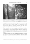
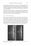
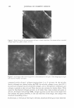
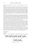
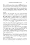
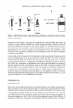
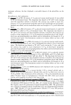
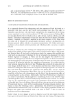
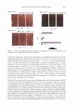
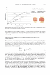
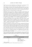
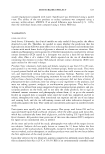
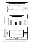
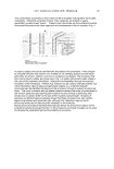
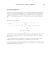
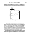
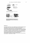
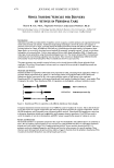
ANALYSIS AND REVIEW OF CELLULITE 385 and more recently by Mirrashed et al. (9) and Smalls et al. (60). High-resolution micro imaging MRI visualizes the architecture of the skin layers at the hypodermal level. It clearly shows the thickness and structural alterations of the connective tissue in both the dermis and subcutaneous adipose tissue (Figure 3). Although it is a very useful method, clinical high-field MRI systems capable of high-resolution imaging are, unfortunately, not yet readily available. Despite these limitations, the method at its current resolution of imaging would dem onstrate a reduction in adipose thickness if an appropriate formulation could be devel oped to deliver actives to the desired site of action. However, since cellulite is clearly about defects in connective tissue structure, MRI would need to be combined with other techniques such as confocal microscopy and histopathology. OPTICAL COHERENCE TOMOGRAPHY (OCT) Optical coherence tomography (OCT) utilizes advanced photonics and fiber optics to obtain images and tissue characterization in human skin. Fully exploited, the technology has the potential to change the way researchers and scientists see and understand the skin. The technology combines the principles of ultrasound with the imaging perfor mance of a microscope. Whereas ultrasound produces images from backscattered sound "echoes," OCT uses infrared light waves that reflect off the internal microstructure within the biological tissues. The frequencies and bandwidths of infrared light are orders of magnitude higher than medical ultrasound signals, resulting in a greatly increased image resolution, 8-25 times greater than that from any other existing modality. Figure 3. Magnetic resonance imaging scans of full-thickness skin (taken from ref. 9). Adipose tissue (b), unlike the dermis (a), is clearly defined.

386 JOURNAL OF COSMETIC SCIENCE Although the resolution of OCT is good, it only extends approximately 500 µm into the skin, and although the epidermis and papillary dermis can be visualized, the subcutis remains obscure. As with ultrasound, the resolution needs to be improved if the tech nology is to be used to visualize subcutaneous adipose tissue at the ultrastructural level and to demonstrate changes in connective tissue structure and organization. LYMPHOGRAPHY This procedure serves to visualize lymph vessels via radiographic examination of the lymphatic system using an injection of contrast medium. If only lymph vessels are to be visualized, water-soluble contrast media are used however, to show the storage pattern of lymph nodes, an oily contrast material is required. Radiological demonstration of lymph vessels and nodes is achieved by direct injection of contrast medium into a peripheral lymph vessel. Several types of needles and/or cannulae are available for cannulation of the lymph vessels, and automatic injectors are used for slow continuous injection of the viscous, oily contrast medium. The water-soluble con trast medium is injected by hand. This technique has never been used in the clinical evaluation of the involvement of lymphatics in the cellulite condition, and before this approach can be considered, clear factual knowledge as to whether cellulite affects lymph draining or causes fibrosclerosis of the lymph vessels is required. This knowledge requirement could be developed using histopathological techniques. DIELECTRIC DEVICES Easily applicable and inexpensive water-specific techniques to evaluate local edema, swollen tissue problems, and fluid retention in humans are not available. In Finland (Delfin Technologies) a recently constructed non-invasive device for local measurement of changes in tissue water in human skin and subcutaneous fat (SSF) was developed (45). The instrument transmits an ultra-high-frequency electromagnetic (EM) wave of 300 MHz into a coaxial line and further into an open-ended coaxial probe that is in contact with the skin. Due to the dimensions of the applied probe, the penetration of the EM field extends to subcutaneous fat. A major part of the EM energy is absorbed by tissue water, while the rest is reflected back into the coaxial line. From the information of the reflected wave, an electrical parameter, directly proportional to the tissue water content, called a dielectric constant of SSF, is calculated. The value increases with increasing water content. The measurement range from normal skin and subcutaneous tissue is from about 15 to 40. The dielectric constant of water is 79 and of adipose tissue about 10 to 20. The measuring depth can be adjusted by changing the dimensions of the probe. The measured dielectric constant is saved as a single value or a mean of three or five samples. This new device enables an easy and non-invasive measurement technique to assess changes of tissue water in SSF. Although the method has yet to be seriously considered and evaluated fully for cellulite studies, it offers excellent potential for demonstrating whether or not water retention and ultimately water structure play a role in the manifestation of the cellulite condition.
Purchased for the exclusive use of nofirst nolast (unknown)
From: SCC Media Library & Resource Center (library.scconline.org)