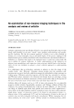
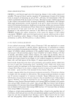
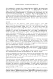
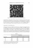
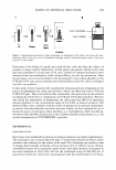
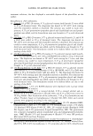
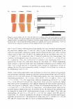
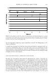
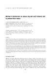
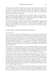
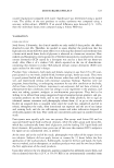
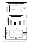
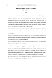
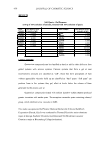
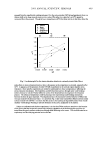
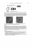
ANALYSIS AND REVIEW OF CELLULITE 383 exercise. However, resolving the fibrosclerotic tissue remains a challenge, as does the increased amount of adipocyte membranous tissue since, although adipocyte content can be reduced, the number of cells is not reduced. Furthermore, understanding why fibro sclerotic tissue forms in this manner needs more in-depth investigation. This can be gleaned from information in studies on breast tissue (51-53) and scar formation and reduction (54,55), as well as from anti-aging studies. As an active endocrine and para crine organ that releases a large number of cytokines that influence inflammation and fibrolysis, increased levels of saturated fatty acids would increase the level of cytokines (adipokines). Consequently, chronic exposure of fibroblasts to cytokines results in fibro sis. Often associated with the cellulite condition are "colorless" striae-striae alba (56), seen as shiny fine lines in the skin. These striae are thought to occur under the pressure of fat accumulation. Seventy percent of females have such striae, including anorexia nervosa patients (12-13 % of the female population). The striae are caused by the presence of more rigid cross-linked collagen, which is easily ruptured under stress. Furthermore, in striae tissue there is an increase in cortisol activity (5 7). LYMPH FLUX Arguments exist in the scientific literature as to the involvement of insufficient lym phatic transport and lymphedema in the cellulite condition, and studies in this area are clearly lacking, giving rise to much controversy (15-17). However, insufficient lym phatic transport in adipose tissue leads to the accumulation of macromolecular proteins and hyaluronan within the extracellular space (58). In lymphedema there is also an accumulation of lymphocytes (macrophages), as well as an increase in the fibroblast, keratinocyte, and adipocyte content of the affected tissue (59). The overgrowth of connective and adipose tissue (also seen in cellulite) in skin and subcutaneous tissue is accompanied by an increase in collagen deposition. EVALUATING THE CONDITION: CLINICAL CONSIDERATIONS There are a number of clinical methods available to evaluate the cellulite condition, though the limitations of each method alone are such that more than one method 1s required in order to correlate clinical findings with any laboratory findings. HISTOP ATHOLOGY The closest work to date as to the real understanding of the cellulite condition is that of Pierard et al. (8). Histopathology tells us that in cellulite superficial fat lobules protrude into the dermis (papillae adiposae), and that there is an unevenness of collagen and elastin fibers. At the site of dimpling, fibrous strands are enlarged, reminiscent of striae distensae (Figure 1). There is also a high content of acid proteoglycans and a2-macroglobulin. There is no tissue dystrophy or lipoatrophy, and the bumpy appear ance is due to a network of connective tissue strands strongly tethering the dermis to the deeper layers, thickened and fibrosclerotic. The limitation of this method is that it is a highly invasive ex vivo technique, and

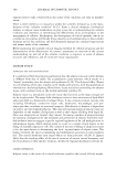
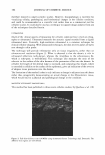
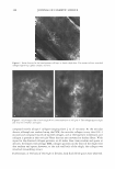
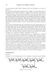
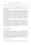
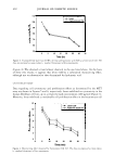
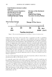
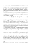
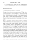
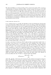
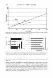
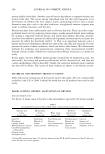
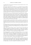
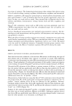
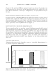
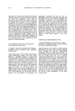
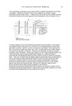
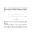
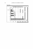
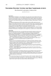
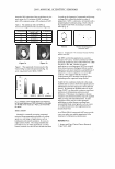
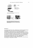
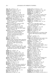
384 JOURNAL OF COSMETIC SCIENCE therefore limited to small scientific studies. However, histopathology is excellent for visualizing cellular, pathological, and biochemical changes in the cellulite condition, and could be recommended as a scientific tool rather than as a commercial tool for cellulite studies. It could also be used as a technique to support image analyses from any of the techniques described herein. ULTRASOUND Much of the clinical aspects of measuring the cellulite condition have relied on echog raphy or ultrasound. Ultrasound measures the acoustic signal recorded from a digital ultrasound wave. Currently, high-resolution ultrasound is a common technique for clinical cellulite imaging. With ultrasound techniques, the skin receives pulses of waves sent through a skin probe. The technique will provide information only on tissue irregularity, rather than on ultrastructural resolution (Figure 2). What is observed is that the dermis is rich in ultrasound reflections (echo-dense), whereas the interphase to the subcutaneous fat, which is echo-poor, is well-defined. This technique also measures the ratio of the subcutis to the surface of the skin because of the protrusion of fat into the dermis. In cellulite the length of the dermal ridge is increased, and comparing this length, which is extended in cellulite to the surface of the epidermis, gives an indication of the severity of adipose tissue protrusion into the dermis. The limitation of the method is that it only indicates a change in adipose tissue thickness rather than categorically demonstrating an actual change in the fibrosclerotic tissue, which would lead to a physical and pathological change in the condition. MAGNETIC RESONANCE IMAGING (MRI) This method has been published in three recent cellulite studies, by Querleux et al. (10) Figure 2. Full-skin-thickness scan (20 MHz) using ultrasound (Cortex Technology ApS, Denmark). The different skin regions are well-defined.
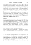
Purchased for the exclusive use of nofirst nolast (unknown)
From: SCC Media Library & Resource Center (library.scconline.org)