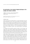
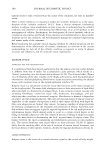
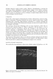
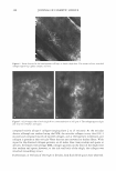
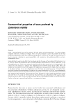
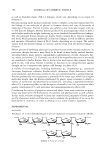
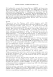
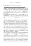
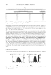
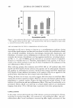
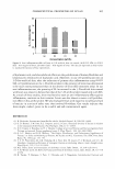
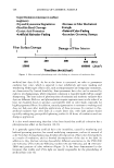
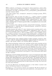
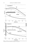
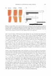
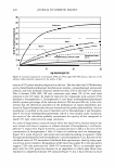
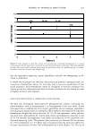
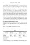
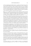
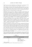
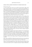
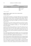
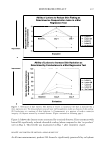
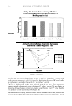
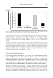
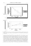
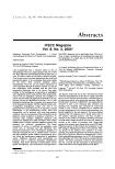
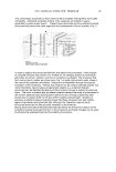
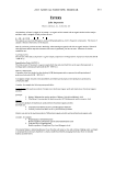
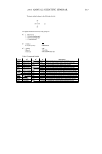
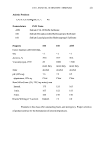
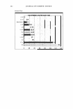
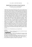
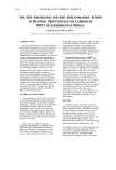
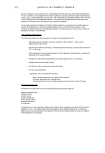
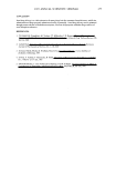
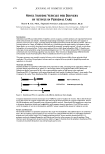
402 110 100 � 90 C .! C 8 80 .! 70 60 50 JOURNAL OF COSMETIC SCIENCE 0 5 10 -+- Levan -o-- Hyaluronic acid 1% 15 Time (hr) 20 25 30 35 Figure 3. Transepidermal water loss (TEWL) of levan and hyaluronic acid (HA) as a function of time. The data are expressed as mean values (± standard deviations) of five experiments. (Figure 4). We observed a trend almost identical to the one listed above. On the basis of these two results, it appears that levan exhibits a substantial moisturizing effect, although not as substantial as that determined for hyaluronic acid. CYTOTOXICITY ASSAY Data regarding cell cytotox1e1ty and proliferation effects as determined by the MTT assay are shown in Figures 5 and 6, respectively. Levan exhibited no cytotoxicity in the human fibroblast cell line, up to a relatively high concentration (100 µg/ml) (Figure 5). Moreover, levan exhibited a considerable cell proliferation effect in the keratinocyte cell 12 -+- Levan 0.2% 10 -o- Hyaluronic acid 0.2% G 8 1 6 G 0 G 4 0 2 0 0 5 10 15 20 25 30 35 Time (min) Figure 4. Moisturizing effect measured by Corneometer CM 825. The data are expressed as mean values (± standard deviations) of five experiments.

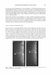
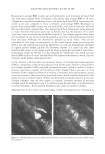
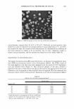
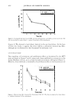
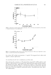
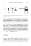
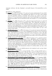
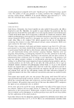
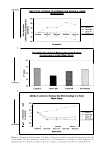
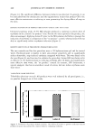
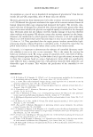
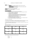
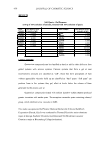
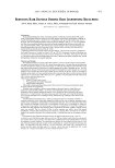
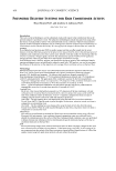
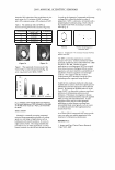
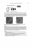
COSMECEUTICAL PROPERTIES OF LEVAN 403 120 115 110 105 � ca 100 · Q) 95 85 80 0 20 40 60 80 100 120 Concentration (ug/ml) Figure 5. Cytotoxicity of levan against human fibroblast cell line. The data are expressed as mean values (± standard deviations) of four experiments. 150 140 130 e... 120 � ca 110 · Q) 100 90 80 70 10 100 1000 Concentration (ug/ml) Figure 6. Cell proliferation effect of levan against human keratinocytes. The data are expressed as mean values (± standard deviations) of four experiments. line-above 30% at high concentrations ( 1 mg/ml). This suggests levan's safety with regard to cosmetic applications. CELL PROLIFERATION TEST WITH 3-DIMENSIONAL ARTIFICIAL SKIN After inducing skin irritation in 3-D artificial skin using 0.05% of SLS, which is used widely as a skin irritant (27), we applied 0.01 mg/ml and 0.05 mg/ml of levan solution, and observed the results. When we compared cell viability in the presence and the absence of levan (SLS only) (Figure 7) we determined that the artificial skin that had been treated with levan exhibited substantially greater cell proliferation (more than a 30% enhancement).
Purchased for the exclusive use of nofirst nolast (unknown)
From: SCC Media Library & Resource Center (library.scconline.org)