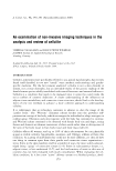
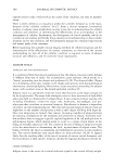
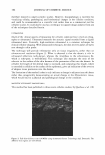
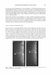
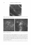
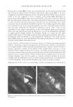
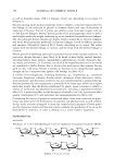
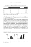
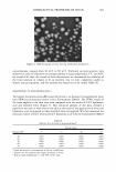
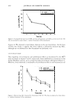
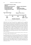
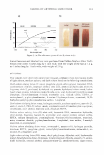
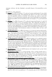
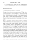
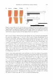
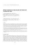
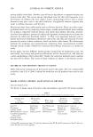
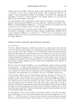
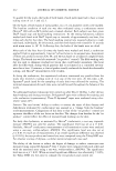
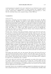
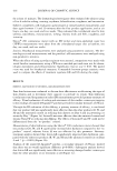
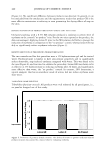
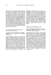
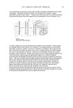
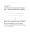
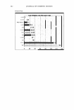
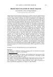
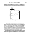
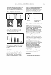
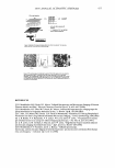
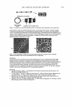
ANALYSIS AND REVIEW OF CELLULITE 387 PRIMOS (FRINGE PROJECTION) PRIMOS is a well known application for measuring changes in skin surface texture and wrinkles. The optical device can also complete 3D measurement of parts of the human body. In instances of measuring an area of 300 x 200 mm, the measurement of the 3D profile is performed with an accuracy of 50 µm. This high accuracy enables application in the cosmetic and medical fields, including dermatology. Furthermore, the technique offers rapid measurements, micron resolution in all axes to ensure the precise determi nation of all measured parameters, highly precise matching of skin areas measured before and after treatment, determination of those characteristics essential for the investigation, and description of body parts with only one instrument. PRIMOS should also detect alterations to the subcutis/cutis structure if this affects the surface topography, i.e., PRIMOS measures the surface irrespective of the causes for changes in skin surface topography. PRIMOS could easily lend application for topical cellulite products devel oped for visualizing surface changes of female thigh skin rather than those products attempting to address the fibrosclerosis or adipose defects. IN VIVO (VIDEO) CONFOCAL MICROSCOPY In vivo confocal microscopy (CFM), using a Vivascope 1500, was employed in a recent study (61) in an attempt to identify specific characteristics of subcutaneous cellulite based on published histopathological findings. The main objective was to compare the impact of cellulite adipose protrusion at the papillary dermis, using in vivo confocal microscopy in the subcutaneous adipose tissue in healthy non-diabetic females, with that of healthy, non-diabetic males. Eleven subjects, five males and six females, were re cruited into the study. Height, weight, and body mass index (BMI) were recorded. All females had visual cellulite on the thighs. In vivo confocal imaging was captured on the front, side, and back aspects of the thighs, 15 images captured per site. Initial results found that striae were seen penetrating into papillary dermis, which were not seen in male skin. Surrounding these "epidermal" striae, which were located and observed within the epidermis at the basal-spinous cell layers, were strands of sclerotic collagen fibers (Figure 4). In contrast, collagen observed in males at the same thigh point was normal-sparse and fine with minimal obvious density (Figure 5 ). In males, imaging before loss of microscope resolution extended to 170 microns, whereas in females this was extended to 200 microns. The average epidermal thickness in males was 5 5 microns compared to 51 microns in females. Larger differences were seen in full-thickness measurements (stratum corneum to the sub-papillary layer of the papillary dermis). However, this may be attributed to loss of resolution in male subjects. Clear differences were observed between females with average BMI and females with greater than average BMI. Females with greater than average BMI had thinner full-thickness measurements (156 microns) than average-BM! females (178 microns). Epidermal thick ness in females with greater than average BMI was less (45.5 microns) than in females with average BMI (62 microns). Collagen as a finely woven meshwork of collagen fibers is found at the papillary layer of the dermis, which includes not only the sub-epidermal papillae situated between the rete ridges but also the sub-papillary layer forming a narrow ribbon between the rete ridges and the sub-papillary blood vessels-papillary dermis. The collagen at this site is

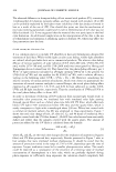
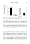
388 JOURNAL OF COSMETIC SCIENCE Figure 4. Striae observed at the basal-spinous cell layer in female thigh skin. The arrows indicate stretched collagen appearing tightly compact and taut. Figure 5. (a,b) Images taken of male thigh skin at same resolution as in Figure 4. The collagen appears light and very fine ("fluffy") and sparse. composed mainly of type I collagen (ranging from 2 to 15 microns). At the reticular dermis, although not resolved using the CFM, the reticular collagen is very thin (0.2-1 micron) and composed mainly of type III collagen, and at the basement membrane zone collagen is present as thin reticular fibers that are not converted to thicker fibers. With regard to the observed collagen patterns, in all males these were random and sparse at all sites. In females with average BMI, collagen patterns on the front of the thigh were also random and sparse however, at the side and back of the thigh, the collagen was stretched (resembling striae). Furthermore, at the back of the thigh in females, dark fluid-filled spaces were observed.
Purchased for the exclusive use of nofirst nolast (unknown)
From: SCC Media Library & Resource Center (library.scconline.org)