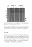
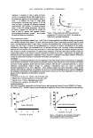
588 JOURNAL OF COSMETIC SCIENCE OZONE AND SKIN: AN 0vERVIEW Giuseppie Valacchi, Ph.D. Center for Comparative Respiratory Biology and Medicine University of California, Davis Davis, CA 95616 The skin, as an interface between the body and the environment, is chronically exposed to stress from both UV radiation and environmental oxidative pollutants such as diesel fuel exhaust, cigarette smoke, halogenated hydrocarbons, heavy metals and 03 (one of the most toxic of these compounds). The skin is protected against oxidative stress by a variety of antioxidants these include enzymatic antioxidants (glutathione peroxidase, superoxide dismutase, catalases) and nonenzymatic low molecular weight antioxidants such as vitamin E isoforms, vitamin C, glutathione (GSH), uric acid and ubiquinol. The distribution of antioxidants in the stratum corneum (SC) follows a gradient with higher concentrations in deeper layers. This may be explained by the fact that SC layers move up in time as a part of the physiological turnover of skin cells and are replaced by freshly differentiated keratinocytes. Therefore, the superficial layer is exposed to chronic oxidative stress for a longer time than the deep layer. Compared with the SC, the surface lipids contain high levels of a- and y tocopherol because of the secretion of vitamin E by sebaceous glands. Eventually, the uppermost layer of the SC will desquamate and the remaining antioxidants and reacted products will be eliminated from the body. It is generally understood that the toxic effects of 03 are mediated through free radical reactions, although 03 is not a radical species per se. They are achieved either directly by the oxidation of biomolecules to give classical radical species (hydroxyl radical) or by driving the radical-dependent production of cytotoxic, nonradical species (aldehydes). Furthermore, the formation of the oxidation products characteristic of damage from free radicals has been shown to be prevented by the addition of the antioxidants vitamin E and C, though the mechanism is not fully understood. The target specificity of 0 3 towards specific compounds together with its physicochemical properties of fairly low aqueous solubility and diffusibility, must be taken into account when a target tissue (lung and skin) is exposed to 03. Within the skin, the SC has been identified as the main target of oxidative damage. As the outer skin barrier, the SC has important functions, limiting transepidermal water loss and posing a mechanical barrier to penetration by exogenous chemicals and pathogens. It comprises a unique two compartment system of structural, non-nucleated cells (corneocytes) embedded in a lipid enriched intercellular matrix, forming stacks of bilayers that are rich in ceramides, cholesterol and free fatty acids. The effects of 03 on cutaneous tissues have recently been evaluated using a murine model (SKHl hairless mice). Skin vitamin E concentration was dramatically reduced after single high dose of 0 3 (10 ppm x 2 h) E. and this was associated with a significant depletion of ascorbate followed by an increase in the lipid peroxidation measured as malondialdehyde (MDA) content. Because its chemical and physic properties, 0 3 does not penetrate through the cells and now it is well accepted the hypothesis that 0 3 mainly reacts within the SC. This hypothesis was supported by further experiments, where hairless mice were exposed to varying levels of 03 for 2 h. Depletion of SC

2007 ANNUAL SCIENTIFIC SEMINAR lipophilic (tocopherols) as well as hydrophilic (ascorbate, urate, GSH) antioxidants was detected upon 0 3 exposure and it was accompanied by a rise in lipid peroxidation measured as 4- hydroxylnonenal (4-HNE) content using both Western blot and immunohistochemical analysis. 589 The further step was to investigate whether the antioxidant depletion and the increase of skin lipid peroxidation products can lead to a cellular active responses and to a modulation of skin pathopshysiology. When hairless mice were exposed for 6 days to 0·8 ppm for 6 h day and the homogenized whole skin was analysed, the increase of proinflammatory markers such as cyclooxygenase-2 (COX-2), nitric oxide synthase (iNOS) and metalloproteinases (MMP's) expression was detected together with markers of cellular stress such as heat shock protein (HSP)32, also known as hame oxygenase-1 (H0-1), HSP 27 and HSP 70. It is therefore possible that bioactive compounds generated by products of 03 exposure may be responsible for the induction a stress insult and the release from the skin tissue of inflammatory markers that can than modulate skin inflammation, skin ageing and wrinkles formation. 03 is also able to modulate skin mouse differentiation and the proliferation measure as increase of proliferating cellular nuclear antigen (PCNA) which is a protein identified as the polymerase associated protein synthesized in the early G1 and S phases of the cell cycle involved in DNA replication and repair and keratin 10 (KlO) which is a protein produced in well differentiated, suprabasal keratinocytes It is not clear how 03 displays its effects, but recent studies have shown that it is able to induce the activation of the transcription factor, NF-KB, by phosphorylation of the kinase, IXBa Finally, in our more recent work on wound-healing we were able to also show that when aged animals (18 months) were exposed to 03 the rate of wound closure was significantly delayed when compared to the young animals. Collectively, our data demonstrate that skin exposure to 03 not only affects antioxidant levels and oxidation markers in the outermost stratum corneum layer, but also induces cellular stress responses in the deeper cellular layers of the skin and this can alter skin physiology.
Purchased for the exclusive use of nofirst nolast (unknown)
From: SCC Media Library & Resource Center (library.scconline.org)